IMAGE

Figure 7

ID
ZDB-IMAGE-230828-91
Source
Figures for Dalla Barba et al., 2023
Image
Figure Caption

Figure 7

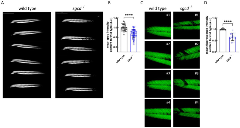
Muscle fiber integrity in the representative wild type and sgcd−/− larvae growing in fish water with 1% methylcellulose added. (A) Representative birefringence images as obtained by viewing zebrafish with a plane polarizing filter. Microscope’s magnification 2.5X. (B) Birefringence quantification of zebrafish larvae as in (A) (wild type N = 39; sgcd−/− N = 45). (C) Representative pictures of the phalloidin staining of whole zebrafish larvae at 5 dpf. Microscope’s magnification 20X. (D) Quantification of the fluorescence intensity of zebrafish larvae stained with phalloidin as in (B) (wild type N = 7; sgcd−/− N = 11). Statistical analysis was performed by the Mann–Whitney test; ****, p ≤ 0.0001.

Acknowledgments
This image is the copyrighted work of the attributed author or publisher, and ZFIN has permission only to display this image to its users. Additional permissions should be obtained from the applicable author or publisher of the image. Full text @ Int. J. Mol. Sci.