IMAGE

Figure 5

ID
ZDB-IMAGE-230828-89
Source
Figures for Dalla Barba et al., 2023
Image
Figure Caption

Figure 5

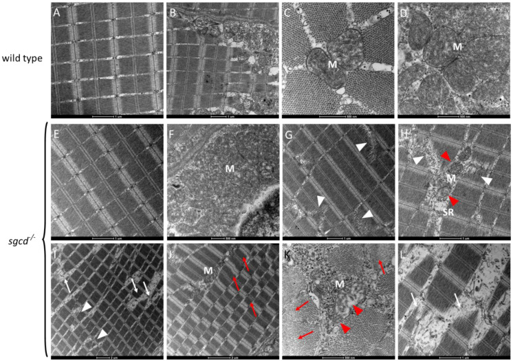
Transmission electron microscopy analysis of the skeletal muscle of the WT and sgcd−/− adult zebrafish lines. Ultrathin sections from 1-year-old WT zebrafish (AD) and age-matched sgcd−/− zebrafish (EL). Scale bar: um, µm. It is possible to observe that in some regions, the contractile tissue and mitochondria (M) of the mutant fish (panel (E,F)) were almost indistinguishable from the wild type (panels (AD)). On the other hand, in most of the sections of sgcd−/− (panels (GL)), clear signs of myopathy were present such as myofibrillar fragmentation (white arrowheads), fiber damage (white arrows), out-of-frame sarcomeric array (panel (J)), hypercontracted fibers (red arrows), and dilated sarcoplasmic reticulum (SR). In these regions, mitochondria are degenerating (panels (H,K), red arrowheads).

Acknowledgments
This image is the copyrighted work of the attributed author or publisher, and ZFIN has permission only to display this image to its users. Additional permissions should be obtained from the applicable author or publisher of the image. Full text @ Int. J. Mol. Sci.