IMAGE

Figure 4

ID
ZDB-IMAGE-230828-87
Source
Figures for Dalla Barba et al., 2023
Image
Figure Caption

Figure 4

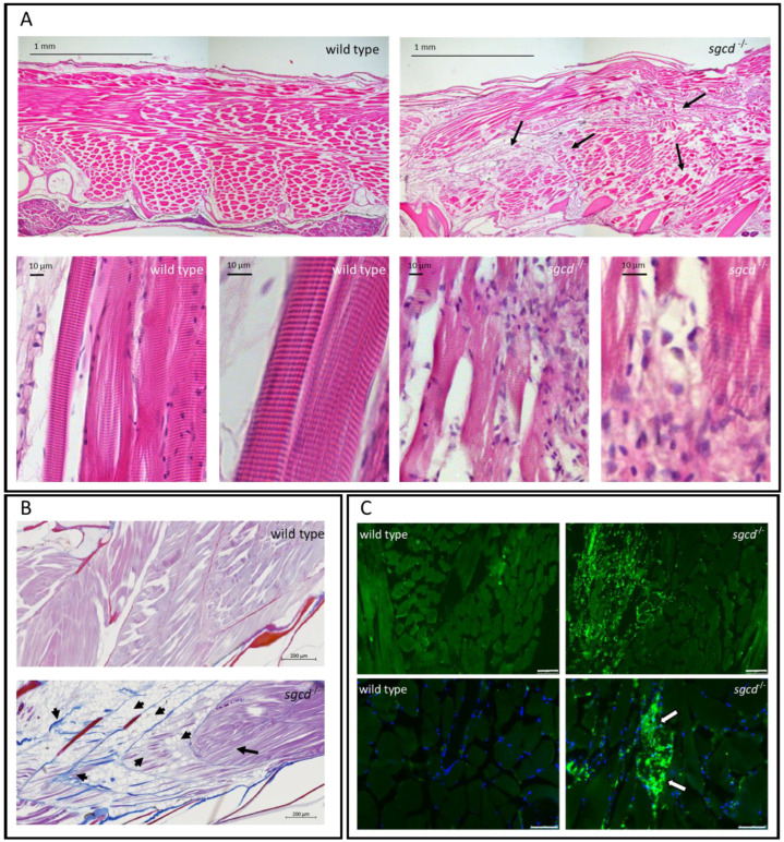
δ-SG deficiency causes defects in the skeletal muscle of adult zebrafish. (A) Hematoxylin and eosin staining of sagittal sections from adult wild type and sgcd−/− zebrafish. The skeletal muscle in the mutated animal was clearly affected, fibers (black arrows) were of different sizes and shapes, and appeared loosely interacting in comparison to the wild type. At high magnification, it is possible to observe that the classical sarcomeric striation, present in wild type tissue, was less evident in the mutant; both fibers and myofibrils were partially disorganized and wavy. Furthermore, a high number of mononucleated cells surrounded the damaged fibers. (B) Azan–Mallory staining of the sagittal section of the wild type and sgcd−/− zebrafish. Arrowheads indicate both the deposition of fibrotic tissue (colored in blue) and the presence of mature adipose tissue replacing the contractile one. The black arrow highlights the presence of a conserved region of skeletal muscle. (C) Immunofluorescence staining with L-plastin (green signal), a pan-leukocytic marker evidenced, at low magnification, the huge number of inflammatory cells that infiltrated the skeletal muscle tissue. Nuclei were counterstained with DAPI (blue signal). At higher magnification, it is possible to observe the L-plastin positive mononucleated cells surrounding a damaged skeletal muscle fiber (arrows). Upper panel bars correspond to 75 µm, lower panel bars to 50 µm Antibody against L-plastin is listed in Table S2.

Acknowledgments
This image is the copyrighted work of the attributed author or publisher, and ZFIN has permission only to display this image to its users. Additional permissions should be obtained from the applicable author or publisher of the image. Full text @ Int. J. Mol. Sci.