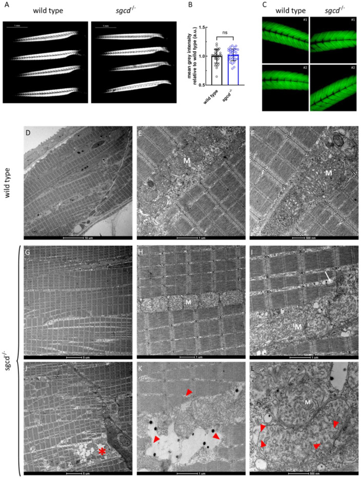
IMAGE

Figure 3

ID
ZDB-IMAGE-230828-86
Source
Figures for Dalla Barba et al., 2023
Image
Figure Caption

Figure 3

Muscle fiber integrity at the macroscopic and ultrastructural level in the sgcd−/− and wild type larvae. (A) Representative birefringence images of the larvae of the two genotypes as obtained by viewing the zebrafish with a plane polarizing filter. Microscope’s magnification 2.5×. (B) Birefringence quantification. Statistical analysis was performed by the Mann–Whitney test; ns, p > 0.05. (C) Representative images of the phalloidin staining of whole zebrafish larvae at 5 dpf. Microscope’s magnification 20×. (DL) Representative ultrathin sections from larvae at 5 dpf. Scale bar: um, µm. The well-preserved array of sarcomeres is evident in both the wild type (DF) and mutated (GI) larvae. In (J), this organization is partially lost, and some fibers are detached from the myosepta (asterisk in (J)), where myofibrillar disarrangement is evident (red arrowheads in (K)). Triad organization in sgcd−/− is almost indistinguishable from that of the wild type (compare (E) with (H)), even if, in a few ultrastructural sections, it is possible to observe that the SR cisternae appeared dilated (white arrow in (I)). Mitochondria (M) appeared normal in both the wild type (E,F) and mutant (H,I) larvae, however, where fibers were damaged, mitochondrial alterations were evident, with detachment of the outer mitochondrial membrane and expansion of the intermembrane space (red arrowheads in (L)).

Acknowledgments
This image is the copyrighted work of the attributed author or publisher, and ZFIN has permission only to display this image to its users. Additional permissions should be obtained from the applicable author or publisher of the image. Full text @ Int. J. Mol. Sci.