IMAGE

Figure 4

ID
ZDB-IMAGE-230828-32
Source
Figures for Staff et al., 2023
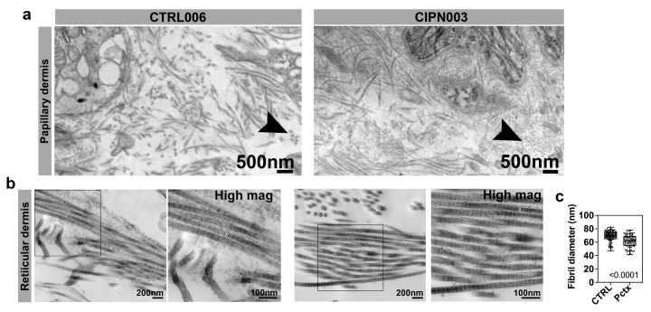
Image
Figure Caption

Figure 4

Dermal collagen abnormalities in CIPN patients. (a) Collagen fibrils in the papillary dermis of a 65-year-old control subject and a 60-year-old patient who was diagnosed with CIPN 35 weeks prior. Cross-sectioned collagen fibrils in the CIPN patient appear granular and small compared with the control subject (black arrowhead). (b) The right panel for each group shows the higher magnification of the boxed area in the left panel. There is no difference in the reticular collagen organization, but the fibrils appear thinner in the CIPN patient, as indicated by white lines in the magnified images. (c) The mean fibril diameter for reticular collagen is significantly reduced in CIPN patients.

Acknowledgments
This image is the copyrighted work of the attributed author or publisher, and ZFIN has permission only to display this image to its users. Additional permissions should be obtained from the applicable author or publisher of the image. Full text @ Cancers