IMAGE

Fig 4

ID
ZDB-IMAGE-230825-24
Source
Figures for Hatzold et al., 2023
Image
Figure Caption

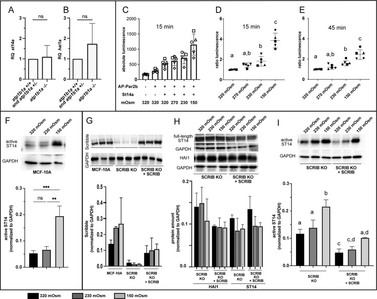
Fig 4 Matriptase activity is increased by hypotonicity and polarity defects.

(A-B) RT-qPCR showing no significant change of st14a and hai1a transcript levels in atp1b1a mutants compared to their siblings (n = 3, cDNA obtained from pools of 15 embryos each at 56 hpf, significances were determined via Student’s t-test; ns, non-significant difference; p>0.05). (C-E) Reporter assay for Matriptase activity towards Par2 cleavage showing that zebrafish Matriptase1a cleaves AP-Par2b more efficiently at lower osmolalities. HEK293 cells were transfected with empty pcDNA3, pcDNA3+AP-Par2b, and pcDNA3+St14a. After 24 hrs in regular / isotonic medium (tonicity of 320 mOsm), cells were exposed to media of 320 mOsm, 270 mOsm, 230 mOsm, and 150 mOsm for 15 and 45 minutes, respectively. (C) At 15 min, absolute luminescence values of AP released into the supernatant progressively increase with increasing hypotonicity / lower tonicity. (D,E) Ratios of luminescence between isotonic and hypotonic media indicate an up to 3.79-fold increase (in 150 mOsm medium) after 15 min (D) and a 2.51-fold increase after 45 min (E). Ratios were determined from values as shown in (C), deducting baseline luminescence (bar 1 in C) from the luminenscences of co-transfected samples obtained at different osmolarities (bars 3–6 in C), normalized against the value at 320 mOsm (isotonic); n = 5, significances were determined via a one-way ANOVA and Tukey’s post hoc test; columns with same superscript letter (a,b,c) are not significantly different (p>0.05). (F) Immunoblot analysis for processed / activated Matriptase-1 (ST14) showing that culturing of MCF-10A cells for 24 hours in hypotonic medium (150 mOsm) leads to a 3.78-fold increase in active endogenous Matriptase-1 compared to cells cultured in isotonic medium (320 Osm). Bar diagram displays mean value of proteins normalized to loading control GAPDH (n = 3); ns, not significantly different (p>0.05); **, ***, significantly different with p<0.01, p<0.001, respectively. (G-H) Scribble knockout (SCRIB KO) in human MCF-10A cells does not affect protein levels of full length ST14 or HAI1. Representative western blots show unaltered amounts of endogenous full-length ST14 (G) and HAI1 (H) proteins in SCRIB-KO cells compared to knockout cells with re-introduced Scribble (SCRIB KO + SCRIB), cultured in media of 320 mOsm, 230 mOsm, or 150 mOsm for 1 hr. Bar diagrams display mean value of proteins normalized to loading control GAPDH (n = 2); all differences are not statistically significant (p>0.05). (I) Loss of Scribble increases active ST14 in media of different osmolalities. Representative western blots showing active ST14 and GAPDH of SCRIB-KO and SCRIB-KO + SCRIB cells cultured in media of 320 mOsm, 230 mOsm, and 150 mOsm for 24 hrs, with highest numbers in cells lacking the epithelial polarity protein Scribble protein and exposed to hypotonic stress. Bar diagram displays mean value of proteins normalized to loading control GAPDH (n = 3). Significances were determined via a one-way ANOVA and Tukey’s post hoc test; columns with same superscript letter (a,b,c,d) are not significantly different (p>0.05).

Acknowledgments
This image is the copyrighted work of the attributed author or publisher, and ZFIN has permission only to display this image to its users. Additional permissions should be obtained from the applicable author or publisher of the image. Full text @ PLoS Genet.