IMAGE

Figure 3

ID
ZDB-IMAGE-230814-221
Source
Figures for Cazorla-Vázquez et al., 2023
Image
Figure Caption

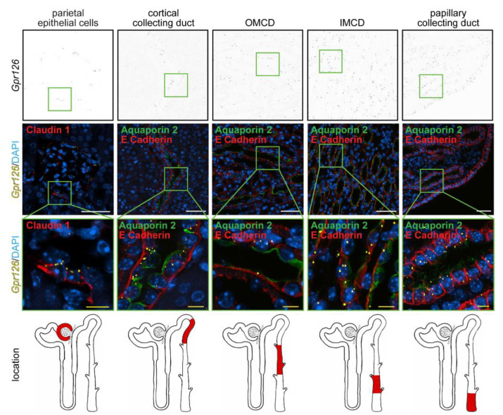
Figure 3

Renal expression of Gpr126 in adult mice. RNAscope® probes targeting the murine Gpr126 mRNA (yellow dots) were combined with renal-specific immunostaining (red) on FFPE kidney tissue of wildtype C57BL/6 mice. Gpr126 signal colocalized the claudin 1-positive parietal epithelial cells lining the Bowman´s capsule and the aquaporin 2/E-cadherin-positive collecting duct. Note Gpr126 expression in the cortical, inner (IMCD), and papillary segments of the collecting duct is enriched compared to the outer segment (OMCD). DAPI was utilized to visualize nuclei. White scale bars: 50 µm. Yellow scale bars: 10 µm.

Acknowledgments
This image is the copyrighted work of the attributed author or publisher, and ZFIN has permission only to display this image to its users. Additional permissions should be obtained from the applicable author or publisher of the image. Full text @ Cells