IMAGE

Figure 1

ID
ZDB-IMAGE-230814-146
Source
Figures for Giardoglou et al., 2023
Image
Figure Caption

Figure 1

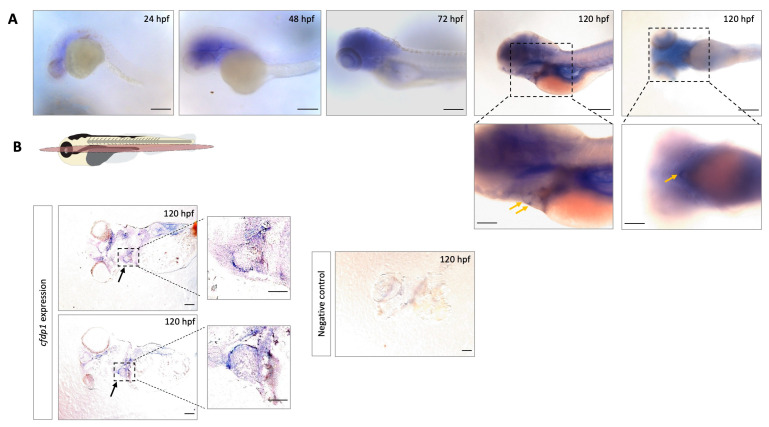
Expression analysis of cfdp1 in different zebrafish development stages shows that cfdp1 emerges during early development. (A) Whole-mount in situ hybridization of cfdp1 in wild-type zebrafish embryos at different development stages. Higher magnification images of the lateral and ventral plane of the 120 hpf cfdp1 ISH-stained embryos are shown in the lower panels, respectively. Arrows point at the region of the heart. The expression of the gene is apparent from 24 hpf and is restricted at the region of the head and the heart by 120 hpf. n = 30 in each of three independent experiments. Scale bar (upper panels) 150 μm; scale bar (lower panels) 200 μm. (B) Top: Illustration of frontal cutting plane of zebrafish; image panels: paraffin sections of 120 hpf ISH-stained embryos with cfdp1 antisense RNA probe (left) and cfdp1 sense RNA (negative control, right). Arrows point at the stained embryonic heart. Scale bar: 50 μm.

Acknowledgments
This image is the copyrighted work of the attributed author or publisher, and ZFIN has permission only to display this image to its users. Additional permissions should be obtained from the applicable author or publisher of the image. Full text @ Cells