
Image
|
Figure Caption
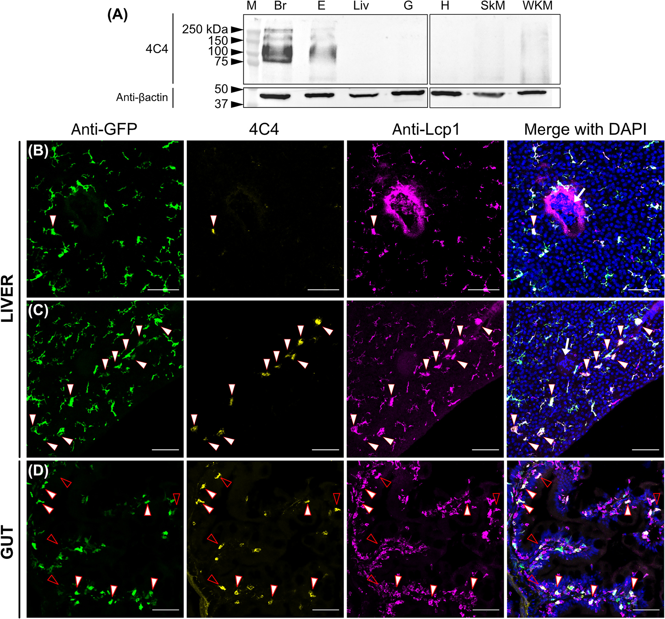
Fig. 6
Expression of lgals3bpb among adult tissues. (A) Tissue distribution by western blot, using the 4C4 antibody and βactin as a loading control. Br, brain; E, eyes; Liv, liver, G, gut; H, heart; SkM, skeletal muscle; WKM, whole kidney marrow (n = 3 fish/tissue). kDa, kilodaltons. (B-D) Immunofluorescence on liver (C,D) and gut (E) sections (14 μm) from an adult Tg(mpeg1:GFP) fish. Anti-GFP (green), 4C4 (yellow), anti-Lcp1 (magenta), and a merge including DAPI staining of the three channels are shown. White arrowheads point to GFP+ 4C4+ Lcp1+ cells while empty arrowheads point to GFP− 4C4+ Lcp1+ cells. White arrow in (C) and (D) merged channels show erythrocyte nuclei indicating the presence of a vessel. Images were taken using a 20× objective and correspond to orthogonal projections. (n = 2). Scale bars 50 μm
Figure Data
Acknowledgments
This image is the copyrighted work of the attributed author or publisher, and
ZFIN has permission only to display this image to its users.
Additional permissions should be obtained from the applicable author or publisher of the image.
Full text @ Dev. Dyn.