
Image
|
Figure Caption
Fig. 5
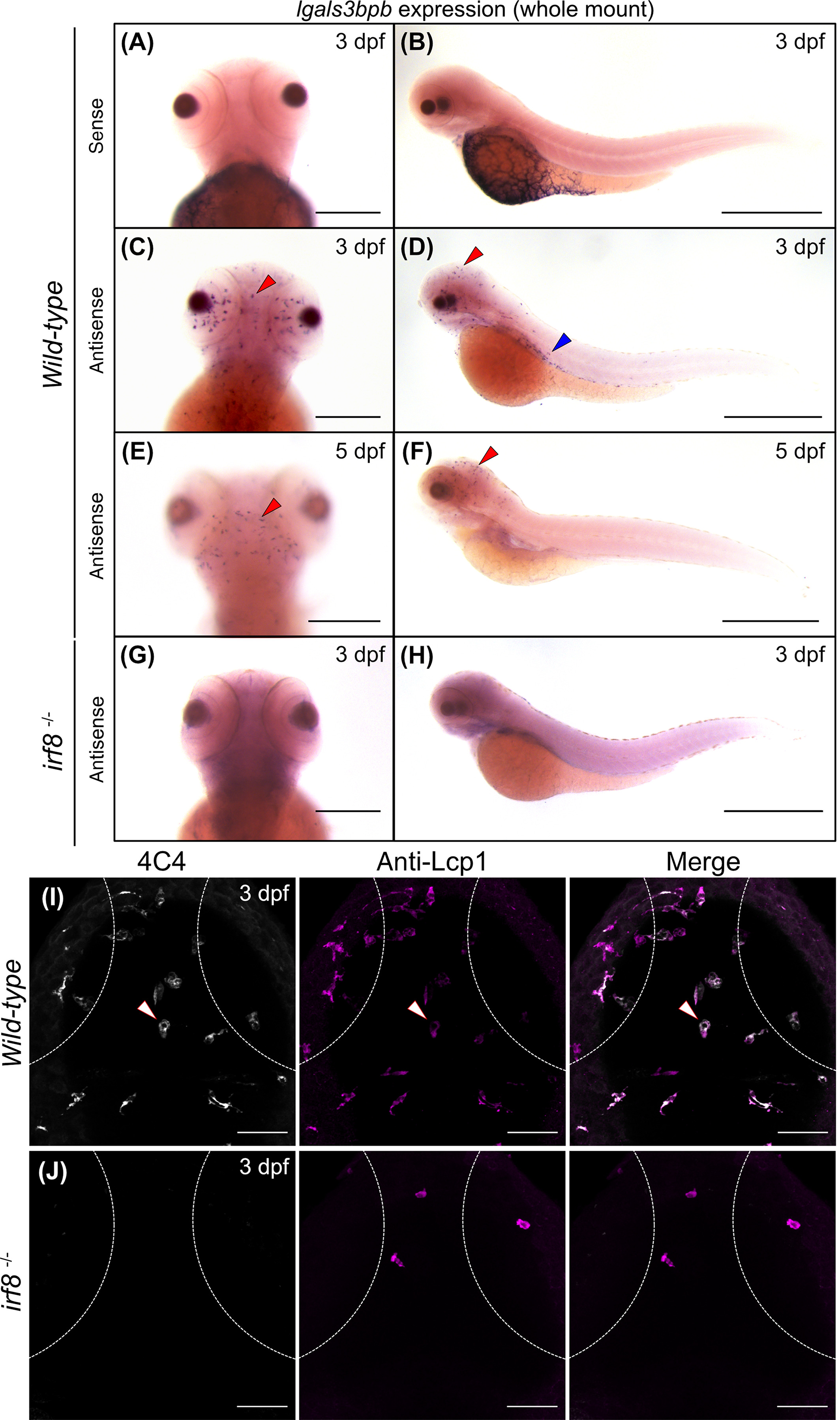
Expression of lgals3bpb in 3 dpf microglia. Whole mount in situ hybridization (WISH) for lgals3bpb sense (A,B) and antisense (C,D) probes in 3 dpf larvae. (C) Dorsal view shows positive staining in the brain in comparison with the sense probe (A). Red arrowheads indicate microglial cells. Scale bar 200 μm. (D) Lateral view shows positive staining in the brain (red arrowhead) as well as in the trunk (blue arrowhead). (E,F) WISH for lgals3bpb in 5 dpf larvae in dorsal and lateral view. Scale bars 200 and 500 μm. (G,H) WISH in irf8−/− mutants at 3 dpf shows absence of lgals3bpb expression in dorsal and lateral view. Scale bars 200 μm and 500 μm. (I,J) Dorsal view of the optic tectum in wild-type and irf8−/− mutants embryos at 3 dpf immunostained with 4C4 (gray) and anti-Lcp1 (magenta) antibodies. Dashed lines represent the eye edges. Images were taken using a 25× water-immersion objective. dpf, days postfertilization
Figure Data
Acknowledgments
This image is the copyrighted work of the attributed author or publisher, and
ZFIN has permission only to display this image to its users.
Additional permissions should be obtained from the applicable author or publisher of the image.
Full text @ Dev. Dyn.