IMAGE

Figure 1

ID
ZDB-IMAGE-230731-103
Source
Figures for Cannone et al., 2023
Image
Figure Caption

Figure 1

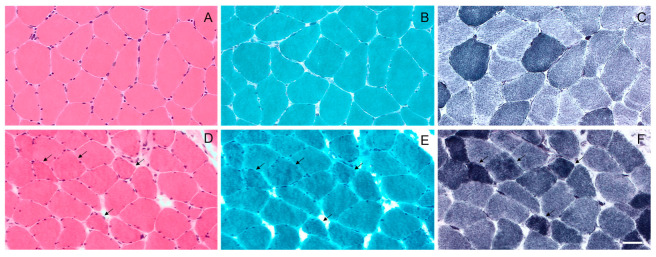
Light microscopy of the muscle biopsy from control subject (AC) and patient with p.Gly154Ser CRYAB mutation (DF). No morphological abnormalities with haematoxylin and eosin (A), modified Gomori trichrome (B) and NADH (C) staining in control subject. Haematoxylin and eosin (D) staining shows fiber size variation in MFM patient. Muscle fibers with amorphous material that stains eosinophilic on haematoxylin and eosin (D) and dark blue on modified Gomori trichrome (E) are also observed (arrows); these fibers have irregular focal areas of decreased or increased NADH staining (F) (arrows). Images were obtained with obj ×20. Scale bar: 50 μm.

Acknowledgments
This image is the copyrighted work of the attributed author or publisher, and ZFIN has permission only to display this image to its users. Additional permissions should be obtained from the applicable author or publisher of the image. Full text @ Int. J. Mol. Sci.