
|
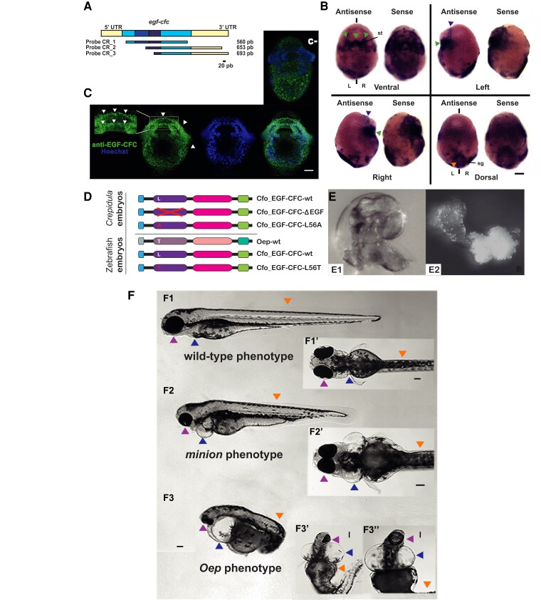
Fig. 4
Cfo-EGF_CFC is not involved in the establishment of LRA in Crepidula fornicata, but a single substitution is able to the rescue of Zebrafish Oep mutants. (A) Cfo-egf-cfc probe cloning designs. Regions in yellow represent the 5′- and 3′-UTRs, and blue corresponds to the coding region. Note that the two darkest fragments correspond to the EGF-like and CFC domains. Probe 1 contains most part of the coding region, whereas probes 2 and 3 contain different lengths of coding region and 3′-UTR. The bar indicates the scale. (B) Localization of Cfo-egf_cfc transcripts by WISH in a C. fornicata preveliger larva with probe 1. The left embryo on each view was incubated with the antisense probe, and the right one corresponds to the sense probe. Green arrowheads point to the symmetrical signal at the stomodeum; purple arrowhead, symmetrical anterior signals; orange, apparent symmetrical shell gland signals. st, stomodeum; sg, shell gland; L, left; R, right; bilateral axis is represented by the vertical bars. Scale corresponds to 30 μm. (C) Ventral view of an immunofluorescent staining for EGF-CFC on a C. fornicata preveliger larva. Green channel shows the staining from a specific antibody designed against this protein, which signal appears to be symmetrical and consistent with the result of WISH (fig. 4BB). Magnification of the stomodeum reveals this protein in the cell membranes (white arrows). The blue channel shows nuclei after Hoechst staining; followed by the merge of both channels. An equivalent larva is provided from the negative control reaction (c−); note the lack of staining at the stomodeum. Scale corresponds to 30 μm. (D) mRNA constructs injected in Crepidula and Zebrafish embryos. (E1) Left side of a wild-type C. fornicata juvenile; (E2) radialized individual resulting from EGF-CFC LOF, after using the construct lacking the EGF-like domain. (F) Zebrafish individuals resulting from the rescue experiment on oep mutants, after injecting zygotes with substitution L56T on the EGF-CFC construct. (F1) Left and (F1′) ventral views of a wild-type phenotype; (F2) left and (F2′) ventral views of a minion phenotype; (F3) left and (F3′ and F3″) ventral views of a classic oep phenotype. Arrowheads point to the structures affected the phenotype: Pink, lens and head; blue, heart; orange, anterior-posterior axis. Scale bars correspond to 100 μm.