IMAGE

Figure 3

ID
ZDB-IMAGE-230630-29
Source
Figures for Stemerdink et al., 2023
Image
Figure Caption

Figure 3

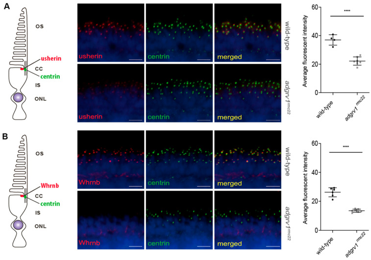
Reduced expression of usherin and Whrnb at the photoreceptor periciliary region of adgrv1rmc22 zebrafish larvae. Retinal cryosections of wild-type and adgrv1rmc22 zebrafish larvae (5 dpf) stained with antibodies directed against usherin (red) (A) or Whrnb (red) (B) and centrin (green). Nuclei are counterstained with DAPI (blue). (A): In wild-type larvae, usherin was present at the photoreceptor periciliary region in close proximity to the connecting cilium marker centrin. The intensity of the usherin signal in adgrv1rmc22 retinal sections was significantly reduced when compared to wild-types (n = 7 adgrv1rmc22 mutant larvae and n = 4 wild-type larvae). (B): In wild-type larvae, Whrnb was present at the photoreceptor periciliary region in close proximity to the connecting cilium marker centrin. The intensity of the Whrnb signal in adgrv1rmc22 retinal sections was significantly reduced when compared to wild-types (n = 7 adgrv1rmc22 mutant larvae and n = 6 wild-type larvae). Intensities of fluorescence signals were quantified (mean ± SD) and plotted in a scatter plot next to the corresponding pictures. **** indicates p < 0.0001 (two-tailed unpaired Student’s t-test). Scale bar: 10 μm.

Acknowledgments
This image is the copyrighted work of the attributed author or publisher, and ZFIN has permission only to display this image to its users. Additional permissions should be obtained from the applicable author or publisher of the image. Full text @ Cells