IMAGE

Figure 1

ID
ZDB-IMAGE-230630-27
Source
Figures for Stemerdink et al., 2023
Image
Figure Caption

Figure 1

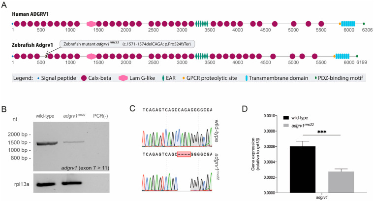
Domain architecture of human and zebrafish ADGRV1 and transcript analysis in homozygous adgrv1rmc22 mutant zebrafish. (A): Schematic representation of human and zebrafish ADGRV1 protein domain structure. Both proteins have a similar repetitive protein domain architecture. Calx-beta: Ca2+-binding calcium exchanger beta domain; Lam G-like: thrombospondin/pentraxin/laminin G-like domain; EAR: epilepsy-associated repeats; GPCR: G protein-coupled receptor; PDZ: post-synaptic density 95, Discs large, Zonula occludens-1 binding motif. (B): RT-PCR analyses of adgrv1 transcripts derived from homozygous adgrv1rmc22 zebrafish larvae (5 dpf) and wild-type siblings revealed a reduction in mutant adgrv1 transcripts when compared to wild-type adgrv1 transcripts. (C): Sanger sequencing confirmed the four-base-pair deletion in the amplicon derived from homozygous adgrv1rmc22 larvae. (D): RT-qPCR analysis of adgrv1 transcripts in wild-type and adgrv1rmc22 zebrafish larvae (5 dpf). Four pools of five larvae per genotype were used in RT-qPCR analysis. *** indicates p = 0.0001 (two-tailed unpaired Student’s t-test).

Acknowledgments
This image is the copyrighted work of the attributed author or publisher, and ZFIN has permission only to display this image to its users. Additional permissions should be obtained from the applicable author or publisher of the image. Full text @ Cells