IMAGE

Figure 4

ID
ZDB-IMAGE-230630-112
Source
Figures for Muñoz-Sánchez et al., 2023
Image
Figure Caption

Figure 4

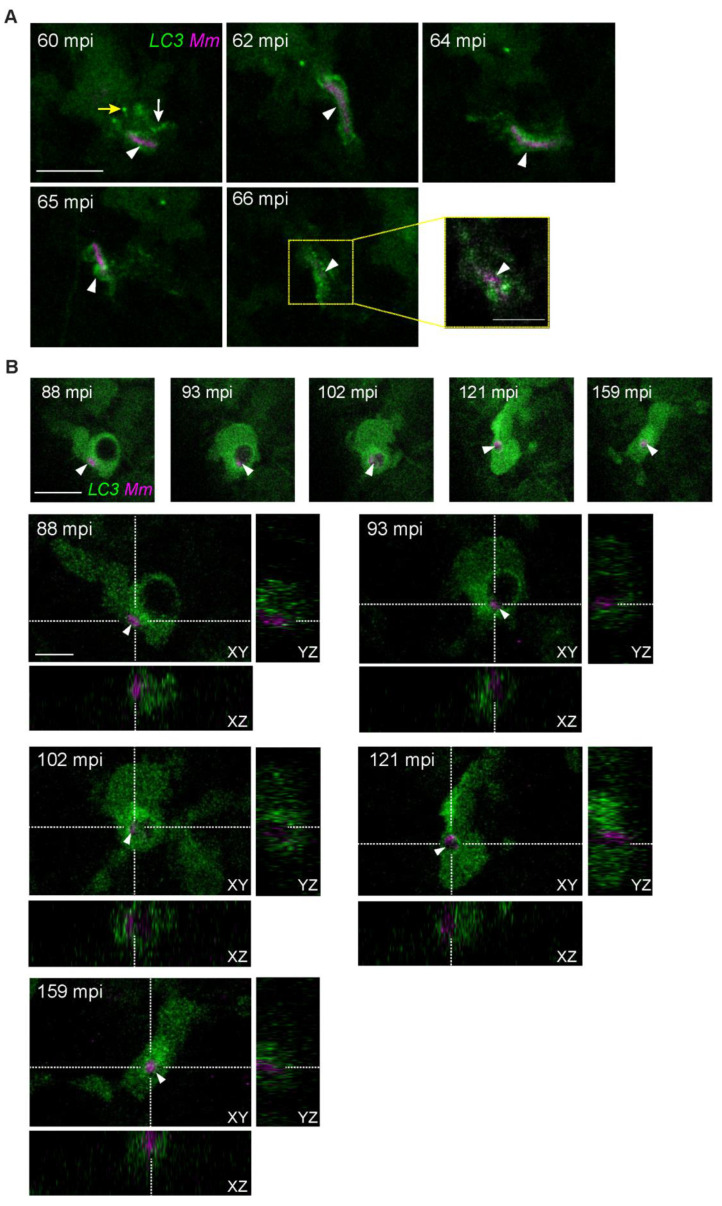
Heterogenous dynamic morphologies of LC3-positive Mm-containing vesicles. Transgenic (CMV:GFP-LC3) zebrafish larvae, labeling LC3 autophagy-related protein (green), were infected with 100 CFU of Mm-mCherry (pseudo color magenta) following the TFI protocol. (A) Maximum projection stills of an infected macrophage that reshapes an LC3-positive Mm-containing vesicle (arrowhead) during migration (60–65 mpi) (Supplementary Video S5). Note how the Mm-containing vesicle (arrowhead) is surrounded by puncta (yellow arrow) and tubular (white arrow) LC3-positive structures and subsequently changes into an elongated form (arrowhead, 62–64 mpi), then reshapes (65 mpi). A zoomed-in Z stack selection of the LC-positive structures at 66 min (outlined yellow) is shown to reveal the presence of Mm. (B) Orthogonal view of the complete Z-stacks, stills capturing the moment when an Mm cluster (white arrowhead) is incorporated into an LC3-positive spacious vesicle (88–93 mpi), after which the vesicle reshapes to a compact form (102–159 mpi) (Supplementary Video S6). Scale bars: 10 μm for all maximum projection stills in A and B, and 5 μm for the zoomed-in area in A and the orthogonal views in B.

Acknowledgments
This image is the copyrighted work of the attributed author or publisher, and ZFIN has permission only to display this image to its users. Additional permissions should be obtained from the applicable author or publisher of the image. Full text @ Biology (Basel)