IMAGE

FIGURE 2

ID
ZDB-IMAGE-230629-89
Source
Figures for Lee et al., 2023
Image
Figure Caption

FIGURE 2

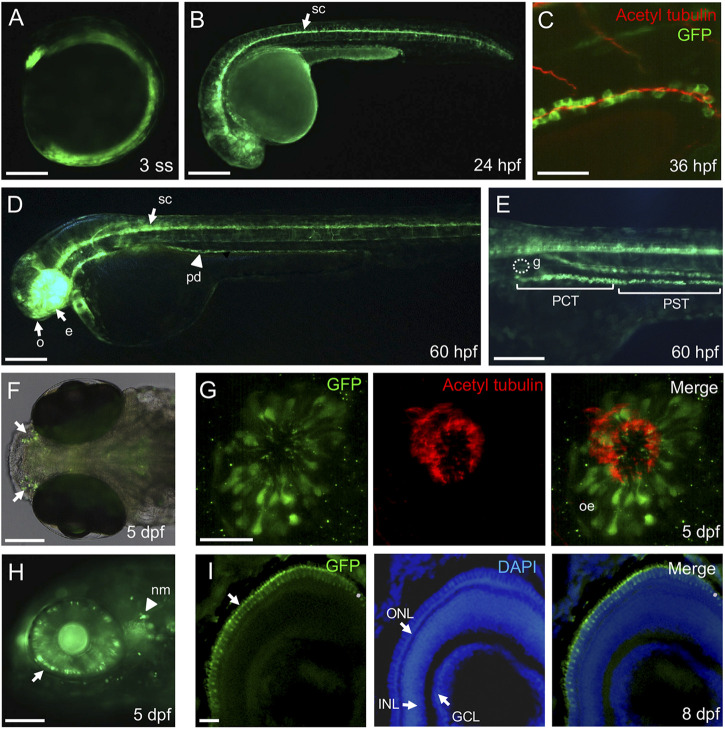
Ciliated cell-specific expression in the stable transgenic IFT46:GAL4-VP16;UAS:mGFP line. (A) Ubiquitous expression of the GFP reporter at three somite-stage embryos. (B) The GFP is strongly expressed in the spinal canal (arrow) at 24 hpf. (C) Immunostaining of anti-acetylated α-tubulin (marking cilia) with GFP in the posterior region of the pronephric duct at 36 hpf. The acetylated α-tubulin signals are overlapped on the apical region of GFP-expressing cells. (D) At 60 hpf, the GFP reporter is expressed in the olfactory region (o), eye (e), spinal canal (arrow), and pronephric duct (arrowhead). (E) High magnification image of the trunk region at 60 hpf. The GFP expression is detected in the proximal convoluted tubule (PCT) and proximal straight tubule (PST) but not in glomerulus (g). (F) Dorsal view of the olfactory region (arrow) in the transgenic IFT46:GAL4-VP16;UAS:GFP line at 5 dpf. (G) Immunostaining of anti-acetylated α-tubulin (marking cilia) with GFP in the olfactory region of the transgenic fish. The olfactory cilia stained with acetylated α-tubulin and GFP reporter detected in the olfactory epithelium (Oe) at 5 dpf. (H) The GFP expression is restricted in the outer nuclear layer of the retina and neuromast cells (nm, arrowhead) at 5 dpf. (I) Transverse section images of the retina at 8 dpf. The GFP reporter is expressed in photoreceptor cells in the outer nuclear layer (ONL) of the retina. Nuclei are counterstained with DAPI. INL, inner nuclear layer; GCL, ganglion cell layer. Scale bars: 200 μm (A,B,D), 100 μm (E,F,H), 50 μm (C), and 25 μm (G,I).

Acknowledgments
This image is the copyrighted work of the attributed author or publisher, and ZFIN has permission only to display this image to its users. Additional permissions should be obtained from the applicable author or publisher of the image. Full text @ Front Cell Dev Biol