IMAGE

Figure 8

ID
ZDB-IMAGE-230629-67
Source
Figures for Mazumdar et al., 2023
Image
Figure Caption

Figure 8

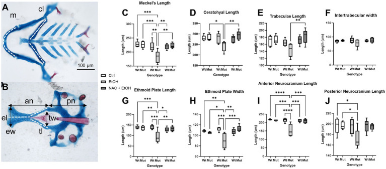
Ethanol-induced craniofacial abnormalities are rescued by concurrent NAC dosage. (A,B) Flatmounts of 5 dpf wildtype craniofacial skeleton displaying the linear measurements used with anterior to the left. (A) Viscerocranium and (B) neurocranium. The cartilaginous elements that were measured: m: Meckel’s length, cl: ceratohyal length, tl: Trabeculae length, tw: Intertrabecular width, el: Ethmoid plate length, ew: Ethmoid plate width, an: Anterior neurocranium length, pn: Posterior neurocranium length. (C–J) Embryo were unexposed, exposed to 1% EtOH from 6–24 hpf, or exposed to 1% EtOH +1 mM NAC from 6–24 hpf. Fish were then grown to 5 dpf for morphometric analyses. Graphs depict craniofacial measurements across each cartilaginous element in all groups (Two-way ANOVA with multiple comparisons, black bars depict range, n = 5 per group, *p < 0.05, **p < 0.01, ***p < 0.001, ****p < 0.0001). In nearly every instance in which ethanol caused a significant reduction in length or width in nnt mutants, the co-exposure with NAC restored the size back to that of wildtype (C,D,G-I). The only exception being posterior neurocranium length, where the co-exposure is not significantly different from wildtype or ethanol-exposed nnt mutants. Ctrl: control, EtOH: ethanol, NAC: N-acetyl Cysteine, Mut: mutant, Wt: wildtype.

Acknowledgments
This image is the copyrighted work of the attributed author or publisher, and ZFIN has permission only to display this image to its users. Additional permissions should be obtained from the applicable author or publisher of the image. Full text @ Front. Neurosci.