
Fig. 2
- ID
- ZDB-IMAGE-230627-2
- Publication
- Flex et al., 2022 - Dominantly acting KIF5B variants with pleiotropic cellular consequences cause variable clinical phenotypes
- All Figures
- Figures for Flex et al., 2022
|
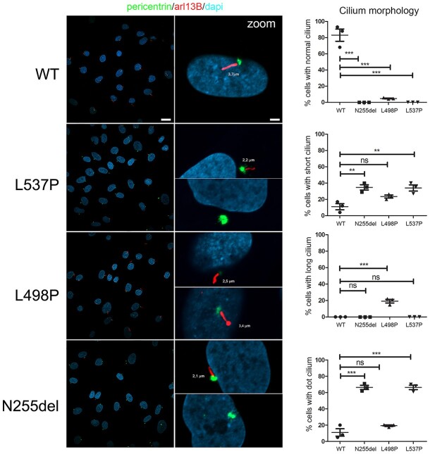
Fig. 2
KIF5B variants cause an aberrant primary cilium morphology. Confocal images showing altered primary cilium morphology in patients’ fibroblasts compared to control cells. Specifically, cilia with altered morphology (either short or characterized by a basal body in absence of any visible cilium [dot cilium], zoomed images) were invariably observed in fibroblasts heterozygous for the variants encoding KIF5BLeu537Pro and KIF5BAsn255del. Similarly, primary fibroblasts carrying the KIF5BLeu498Pro protein showed a large proportion of cells having cilia altered in length and thickness (zoomed images). Primary cilia are labeled with ARL13B (red), basal bodies and nuclei are labeled with pericentrin (green) and DAPI (blue), respectively. Scale bars are respectively 10 μm (left) and 2 μm (right). Cells were analyzed for each line over three independent experiments (50 cells/line each) for a total of 150 cells/line scored. P values were calculated by one-way ANOVA with Tukey’s correction for multiple testing. Graph bars show mean ± SEM.