
|
Fig. 3
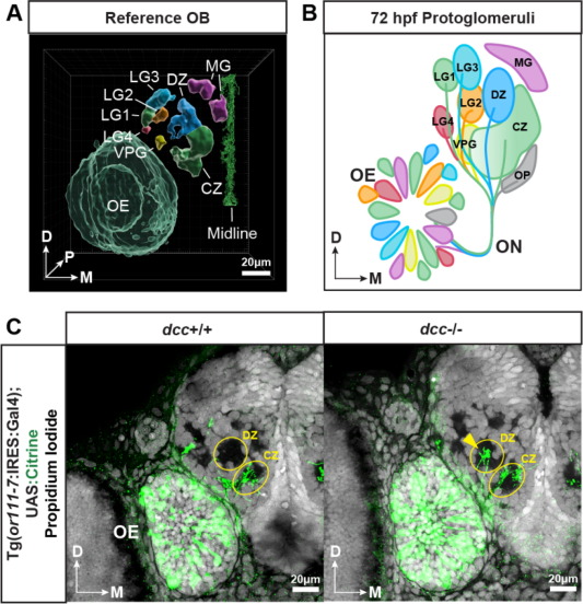
Fig. 3. Protoglomerular identity and targeting. (A) Model of a 72 hpf reference OB hemisphere including the OE (left) and the midline (right). Individual protoglomeruli, the OE, and midline structures were segmented from an unbiased consensus template created from twenty OB hemispheres. (B) Schematic of the 72 hpf zebrafish olfactory system as originally inferred by OMP:RFP and TRPC2:Venus expression. (C) Representative confocal sections of wild type and dcc mutant siblings showing Tg(or111-7:IRES:Gal4);UAS:Citrine expressing OSN axons (green) terminating in individual protoglomeruli. Yellow arrow indicates misprojecting axons into the DZ protoglomerulus.