
|
Figure 1
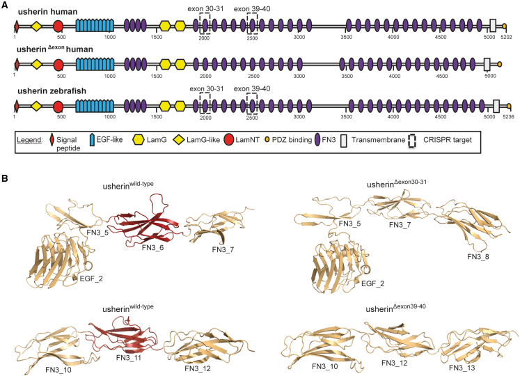
In silico modeling of usherin domain architecture after USH2A exon 30–31 and exon 39–40 skipping
(A) Schematic representation of the domain architecture of the large isoform (isoform B) of human and zebrafish usherin. Isoform B of human and zebrafish usherin are composed of the same repetitive domain architecture that includes a signal peptide, a laminin G-like domain (LamG-like), a laminin N-terminal domain (LamNT), 10 EGF-like motifs, four fibronectin type III (FN3) domains, two laminin G domains (LamG), 28 additional FN3 domains, one transmembrane domain, and a short intracellular region with a C-terminal class I PDZ binding motif. Skipping of exons 30–31 and exons 39–40 both result in the absence of one FN3 domain. The domains that are lost in human and zebrafish usherin are indicated with dashed boxes. Both proteins that result from exon skipping are composed of the same protein domain architecture (visualized as usherinΔexon). Numbers indicate amino acids. (B) For both skipping of USH2A exons 30–31 and exons 39–40, 3D homology modeling predicts the removal of exactly one FN3 domain without disturbing the folding of neighboring FN3 domains. The part of usherin that is encoded by the targeted exons is depicted in red.