IMAGE

Fig. 4

ID
ZDB-IMAGE-230613-31
Source
Figures for Čapek et al., 2023
Image
Figure Caption

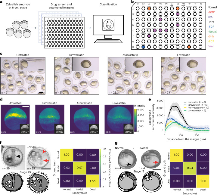
Fig. 4 Applications of EmbryoNet in drug screening and other species.

a,b, Automated phenotype-based drug screening. a, Schematic of the phenomic drug screen. Embryos were exposed to compounds in 96-well plates and imaged for 24 h. Phenotypes were classified automatically by EmbryoNet. b, Layout of BML-2843 library plate 2 with majority phenotype classification for each well. Simvastatin in well H-02 was classified as −FGF. c, Statins identified by EmbryoNet in the drug screen caused body axis defects similar to −FGF loss-of-function phenotypes. d, Representative immunofluorescence images of the FGF signaling transducer pErk in untreated and statin-treated embryos, respectively. The representative images have pErk profiles that are closest to the mean signaling profile of each group. Images are shown at the same contrast and brightness. The inserted lower panels show cell nuclei labeled with DRAQ7. e, Quantification of background-subtracted pErk fluorescence intensity gradients in wild-type (black), simvastatin-treated (blue), atorvastatin-treated (yellow) and lovastatin-treated (green) embryos along the marginal-to-animal pole axis. The error envelopes show s.e.m. f,g, Extension of EmbryoNet to other species. f, Images of wild-type (left) and Nodal-deficient (right) medaka embryos with the confusion matrix of classification performance. g, Images of wild-type (left) and Nodal-deficient (right) three-spined stickleback embryos with the confusion matrix of classification performance. Black arrows point to somites in healthy embryos, while red arrows point out missing somites. The red arrowhead shows a mispatterned central nervous system. Scale bars: 500 µm (c,f,g) and 200 µm (d).

Acknowledgments
This image is the copyrighted work of the attributed author or publisher, and ZFIN has permission only to display this image to its users. Additional permissions should be obtained from the applicable author or publisher of the image. Full text @ Nat. Methods