IMAGE

Figure 6.

ID
ZDB-IMAGE-230606-25
Source
Figures for Hamling et al., 2023
Image
Figure Caption

Figure 6.

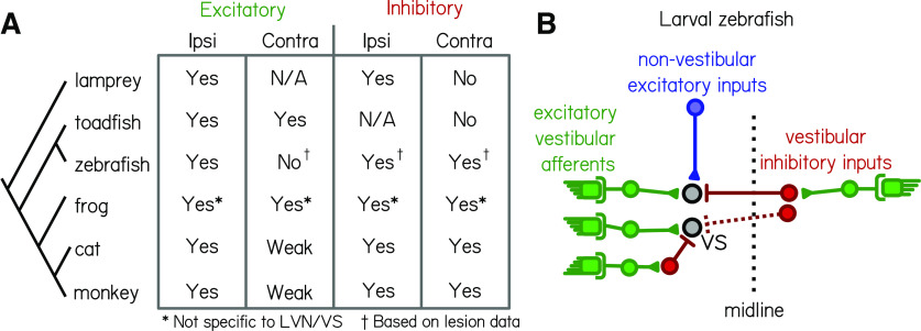
Comparative synaptic architecture of zebrafish vestibulospinal neurons. A, Summary of previous circuit mapping of functional synaptic connections between vestibular afferents and secondary vestibular neurons across species [lamprey (Rovainen, 1979); toadfish (Korn et al., 1977); zebrafish (Liu et al., 2020; current study); frog (Ozawa et al., 1974; Holler and Straka, 2001; Pfanzelt et al., 2008; Malinvaud et al., 2010); cat (Shimazu and Smith, 1971; Uchino et al., 1999, 2001; Kushiro et al., 2000; Ogawa et al., 2000); and monkey (Goldberg et al., 1987; Highstein et al., 1987)]. All characterizations were from vestibulospinal neuron homologs, except for the frog (asterisk) where data were not specific to vestibulospinal neurons in the lateral vestibular nucleus. Connections were determined by afferent activation, except where only afferent lesion data from the current study were available (dagger). B, Vestibulospinal neurons (“VS”, black circles) receive convergent high-amplitude excitatory inputs (green) from irregular afferents originating with the ipsilateral utricle (see also Liu et al., 2020), low-amplitude excitatory inputs (blue) from extravestibular sources and inhibitory inputs (red) from either the ipsilateral or contralateral utricle.

Acknowledgments
This image is the copyrighted work of the attributed author or publisher, and ZFIN has permission only to display this image to its users. Additional permissions should be obtained from the applicable author or publisher of the image. Full text @ eNeuro