
Figure 6
- ID
- ZDB-IMAGE-230501-153
- Publication
- Pose-Méndez et al., 2023 - Lifelong regeneration of cerebellar Purkinje cells after induced cell ablation in zebrafish
- All Figures
- Figures for Pose-Méndez et al., 2023
|
Figure 6
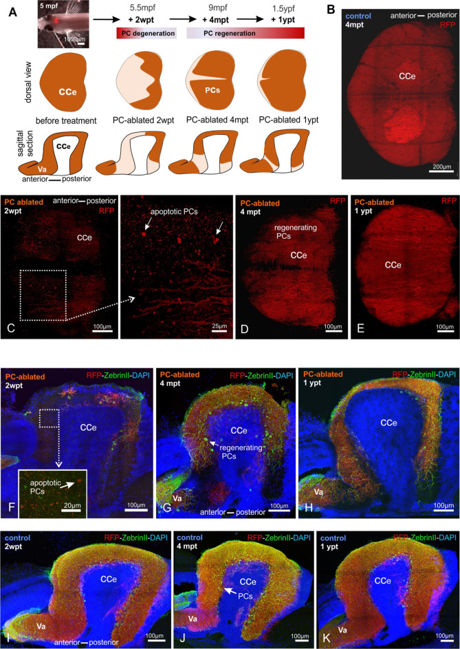
(A) In vivo stereomicroscopy showing tagRFP-T fluorescence in the PC layer, and illustration of the time course of fluorescence recovery after PC ablation in adults (at 5 mpf) monitored until 1 ypt. Representative confocal images of whole mount cerebelli from dorsal view (B–E) and sagittal vibratome sections after immunostaining with the antibodies anti-tagRFP and anti-ZebrinII (F–K), comparing the cerebellum in ablated fish (endoxifen treated; C–E, F–H) vs control group (dimethyl sulfoxide [DMSO] treated; B, I–K). Arrows in C, F point out apoptotic bodies. Equivalent results were observed in two additional independent ablations in adult cerebelli (data not shown).
Monitoring of the Purkinje cell (PC) layer after induced PC ablation in adults.