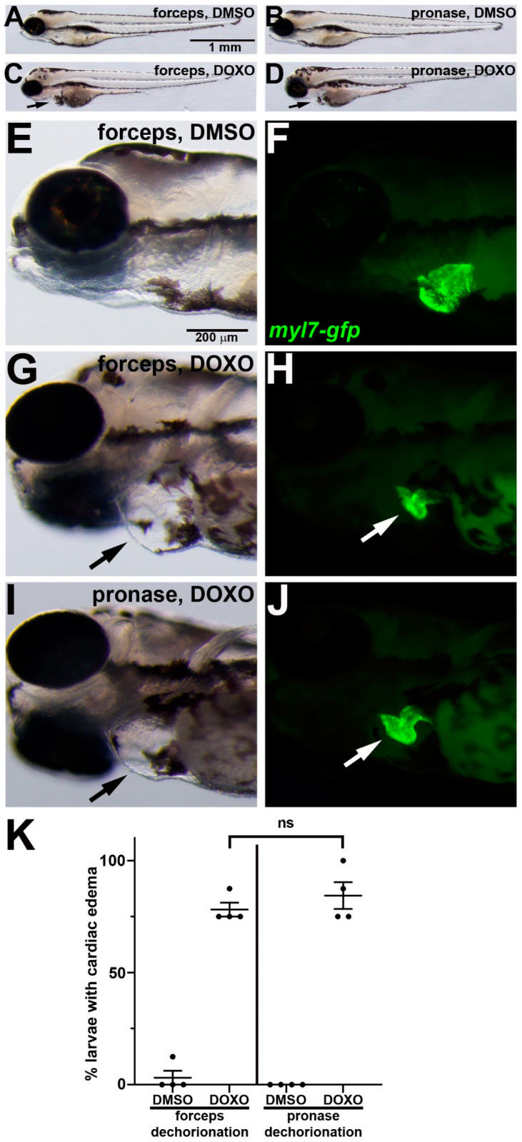
IMAGE

Figure 4

ID
ZDB-IMAGE-230427-5
Source
Figures for Hasegawa et al., 2023
Image
Figure Caption

Figure 4

DOXO-induced cardiotoxicity in zebrafish embryos following manual or pronase dechorionation. (AJ) 96 hpf larvae that were treated from 24 hpf with (A,B,E,F) DMSO or (C,D,GJ) DOXO. Lateral views of larvae show anterior to the left. (EJ) Magnifications of head and cardiac region show paired brightfield and fluorescent images. Black arrows in (C,D,G,J) point to swelling around the heart. In (F,H,J), myl7-gfp (green) labels the myocardium, which is reduced in DOXO treatment ((H,J), white arrows). (K) Graph of % larvae showing cardiac edema. n = 4 treatment replicates, each consisting of eight embryos. ns = not significant.

Acknowledgments
This image is the copyrighted work of the attributed author or publisher, and ZFIN has permission only to display this image to its users. Additional permissions should be obtained from the applicable author or publisher of the image. Full text @ J Dev Biol