
|
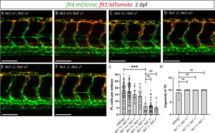
Figure 6.
(A–F) Confocal images of blood and lymphatic vasculature in the trunk of 2 dpf old embryos derived from tie1; tie2 double heterozygous parents, showing no genetic interaction between tie1 and tie2. (G) Quantification of PLs at 2 dpf and of thoracic duct fragments at 5 dpf (siblings: n = 50; tie1+/−; tie2+/−: n = 62; tie1+/+; tie2−/−: n = 13; tie1+/−; tie2−/−: n = 20; tie1−/−; tie2+/+: n = 10; tie1−/−; tie2+/−: n = 32; tie1−/−; tie2−/−: n = 10). (H) TD fragments were counted over the anterior-most 10 somites (siblings: n = 47; tie1+/−; tie2+/−: n = 34; tie1+/+; tie2−/−: n = 5; tie1+/−; tie2−/−: n = 16). Mann–Whitney test was applied for statistical analysis. ***p = 0.0002, ns = not significant. Scale bar = 100 µm. dpf, days post-fertilization; PL, parachordal lymphangioblast; TD, thoracic duct.
tie1; tie2 double mutants show no exacerbation of the tie1 mutant defects.