
Image
|
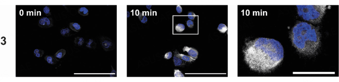
Figure Caption
Figure 4
Intracellular localization of 3 (25 µM) using melanoma cells (518A2) after 10 min. The uptake was visualized using a Cu(I)-catalyzed reaction with 3-azido-7-hydroxycoumarin (white), and the nuclei were counterstained (Nuclear Green, blue). The right image shows the magnified section marked with a white box. The experiment was carried out in duplicate. The scale bar corresponds to 100 µm (left) or 25 µm (right), magnification of 630×.
Acknowledgments
This image is the copyrighted work of the attributed author or publisher, and
ZFIN has permission only to display this image to its users.
Additional permissions should be obtained from the applicable author or publisher of the image.
Full text @ Cancer Drug Resist