
|
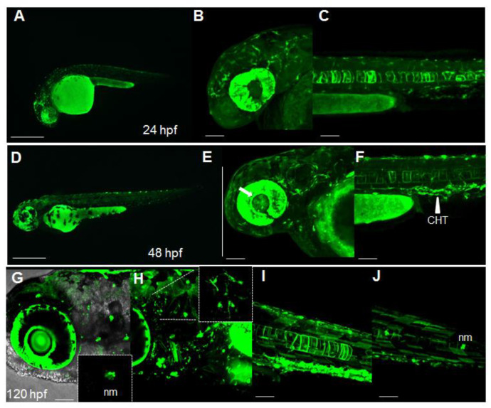
Figure 1
Dynamic spatiotemporal reporter expression in the Nrf2/ARE transgenic line. (A) Whole-mount fluorescence microscopy of a Tg(8xAORE:EGFP)ia201 larva 24 hpf, showing the widespread expression of reporter-expressing cells in both cephalic and more caudal regions. (B,C) Confocal Z-stack projection of the cephalic area (B) and trunk region (C) of a 24 hpf transgenic larva in which strong fluorescent cells are detectable in the brain, eye and notochord. (D) Whole-mount fluorescence microscopy of a 48 hpf Tg(8xAORE:EGFP)ia201 larva, showing the prolonged expression of the reporter in the cephalic and trunk regions. (E,F) Confocal Z-stack projection of the cephalic area (E) and trunk regions (F) of the same 48 hpf transgenic larva exhibiting novel expression domains in the lens and inner eye (white arrow) and the caudal hematopoietic tissue (CHT) (white arrowhead). (G–J) Confocal Z-stack projection of the cephalic area (G,H) and trunk regions (I,J) of a 120 hpf transgenic larva showing fluorescent neuromasts (nm) and protrusion-carrying brain cells (small inset in H), as well as muscle fibers along the trunk (I,J). All images are lateral views with anterior to the left. Scale bar in (A,D): 500 μm; in (B,C,E,F) and (G–J): 100 μm.