
|
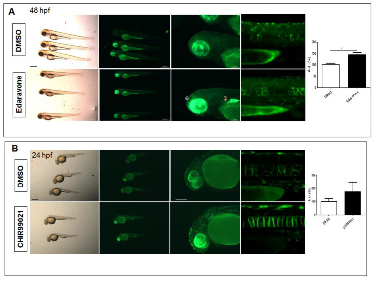
Figure 3
Pharmacological testing of Nrf2/ARE reporter fish. (A) Whole-mount bright field and fluorescence microscopy acquisition of 48 hpf transgenic larvae treated with DMSO or the radical scavenger Edaravone for 24 h. An evident increase in fluorescence is detected in the eye (e) and gut (g) of Edaravone-treated larvae when compared to DMSO-treated control fish. (B) Whole-mount bright field and fluorescence microscopy acquisition of 24 hpf transgenic larvae treated with DMSO or the GSK3β antagonist, CHIR99021, for 16 h. Increased reporter fluorescence is observed throughout the whole larvae when compared to the DMSO-treated control fish. The magnifications of the trunk regions are confocal Z-stack acquisitions. The graphs reported on the right depict the ImageJ-based quantification of the selected trunk region of 5 independent treated fish. (* p < 0.05, t-test) All images are lateral views with anterior to the left. Scale bars in the panels with multiple fish: 500 μm; scale bars on the panels with a single magnified larva: 100 μm.