
|
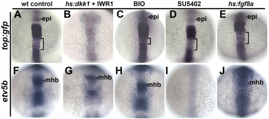
Fig. 1 Early responses to modulation of Wnt and Fgf. (A–J) Dorsal views (anterior to the top) showing expression at 14 hpf of the artificial Wnt reporter top:gfp (A–E) and the Fgf-target gene etv5b (F–J) following treatment at 12 hpf with DMSO (control), hs:dkk1 (39 °C for 1 h) + 10 μM IWR-1 added immediately after heat shock, BIO (5 μM), SU5402 (70 μM), or hs:fgf8a (39 °C for 1 h). Epiphysis (epi) and midbrain-hindbrain border (MHB) are indicated. Brackets mark a zone of weaker top:gfp expression in the hindbrain. The same conditions were used in subsequent figures, except for changes in treatment duration and time of fixation as noted.
Reprinted from Developmental Biology, 492, Tan, A.L., Mohanty, S., Guo, J., Lekven, A.C., Riley, B.B., Pax2a, Sp5a and Sp5l act downstream of Fgf and Wnt to coordinate sensory-neural patterning in the inner ear, 139-153, Copyright (2022) with permission from Elsevier. Full text @ Dev. Biol.