
Image
|
Figure Caption
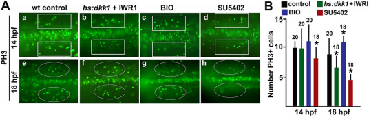
Fig. 4 Effects on proliferation. (A) Dorsal view of embryos stained for phospho-histone H3 to label mitotic cells at 14 and 18 hpf under conditions indicated at the top of the panel. Marquees around the otic placode (200 μm × 50 μm rectangles) or otic vesicle (ovals) were used for quantitation. Ovals delimit the edges of the otic vesicle. (B) Mean number of cells stained with anti-PH3 inside the marquees shown in (A) at 14 and 18 hpf.
Figure Data
Acknowledgments
This image is the copyrighted work of the attributed author or publisher, and
ZFIN has permission only to display this image to its users.
Additional permissions should be obtained from the applicable author or publisher of the image.
Reprinted from Developmental Biology, 492, Tan, A.L., Mohanty, S., Guo, J., Lekven, A.C., Riley, B.B., Pax2a, Sp5a and Sp5l act downstream of Fgf and Wnt to coordinate sensory-neural patterning in the inner ear, 139-153, Copyright (2022) with permission from Elsevier. Full text @ Dev. Biol.