
|
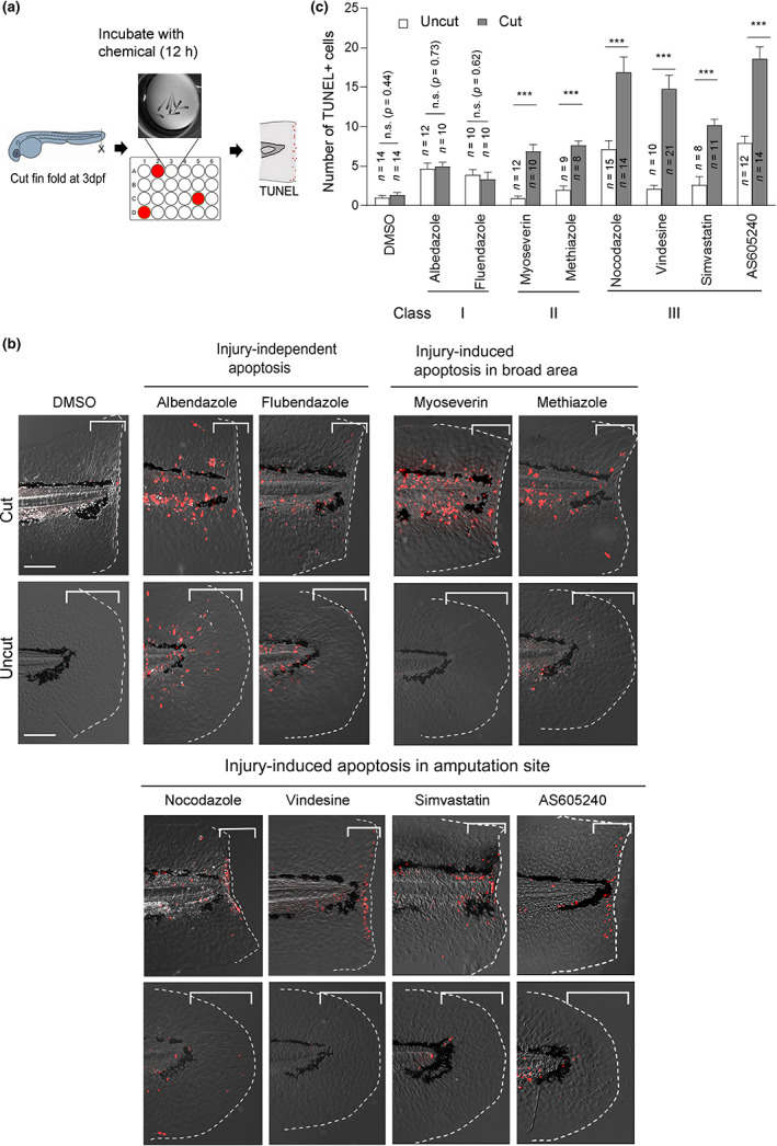
Fig. 1
Screening compounds that induce apoptosis in the larvae fin fold. (a) Schematic overview of the screening procedure. Wild type (WT) zebrafish larvae were amputated at 3 days post fertilization (dpf) and incubated with the compounds for 12 h in a 24‐well plate. Uncut larvae were also incubated as a control. Apoptotic effects were evaluated by TUNEL (terminal deoxynucleotidyl transferase dUTP nick end labeling) staining. (b) Representative apoptosis phenotypes caused by the respective chemicals. Class I compounds (albendazole and flubendazole) induced ubiquitous apoptosis in cut and uncut larvae. Class II and III compounds induced apoptosis in response to tissue amputation. Class III compounds (nocodazole, vindesine, simvastatin, and AS605240), induced apoptosis mostly close to the amputation site, whereas class II compounds (myoseverin and methiazole) induced apoptosis in broader areas. Scale bar, 50 μm. (c) Quantification of the number of apoptotic cells in the bracketed areas from the posterior end of the notochord to the amputation plane in (b). Data are presented as the mean ± SEM. Student's t‐test. ***p < 0.001. n.s., not significant. DMSO, dimethyl sulfoxide