
|
Fig. 1
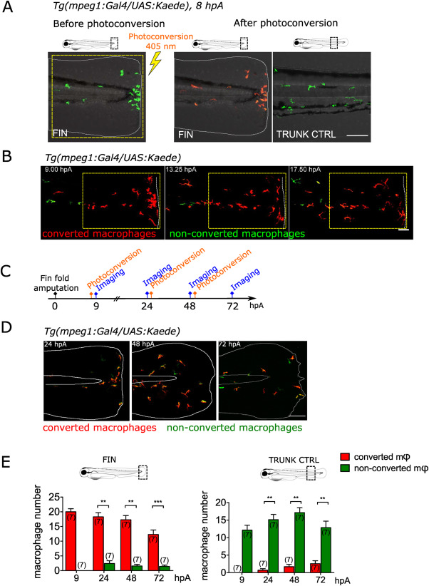
Fig. 1. Same population of macrophages is present at the wound during healing and repair (A) The caudal fin fold of Tg(mpeg1:Gal4/UAS:Kaede) larvae was amputated at 3 dpf. Recruited Kaede+ macrophages (green) were photo-converted to red fluorescence using UV at using 403 nm laser illumination in the wound region at 8 hpA. Representative confocal images of the fin fold before the photoconversion (left) and after the photoconversion (middle). On the right, control image of the trunk region taken after the photoconversion, outside of the wound area showing non-photoconverted macrophages (green). Representative merged images of the Kaede fluorescence in the green and red channels and bright field images. Yellow dashed line indicates the photoconverted area. The white lines outline the fin fold. Scale bar: 100 μm. (B) Representative frames (Maximum projection) of the time lapse imaging of photoconverted area, spanning from 9 to 19 hpA, with a time step of 10 min. White dashed line indicate the wound margin. Scale bar: 50 μm. (C) Schedule of the photoconversion pulse-chase experiment. Fin folds of Tg(mpeg1:Gal4/UAS:Kaede) larvae were amputated at 3 dpf. Photoconversions of Kaede+ macrophages were performed at 8, 25 and 49 hpA. Images were acquired at 9, 24, 48 and at 72 hpA. (D) Representative maximum projections of fin folds at 24, 48 and 72 hpA, taken before the new photoconversion; Merged images of the Kaede fluorescence (green and red channels). The white lines outline the fin fold and the notochord. Scale bar: 100 μm. (E) Quantification of photoconverted (red) and non-photoconverted (green) macrophages (Φ) at the wound (left) and in control trunk region (right), in indicated time point. Two independent experiments, number of larvae is indicated in the figure, Mann-Whitney test, two-tailed, **p < 0.01, ***p < 0.001. (For interpretation of the references to colour in this figure legend, the reader is referred to the Web version of this article.)
Reprinted from Free radical biology & medicine, 192, Sipka, T., Park, S.A., Ozbilgic, R., Balas, L., Durand, T., Mikula, K., Lutfalla, G., Nguyen-Chi, M., Macrophages undergo a behavioural switch during wound healing in zebrafish, 200-212, Copyright (2022) with permission from Elsevier. Full text @ Free Radic. Biol. Med.