
|
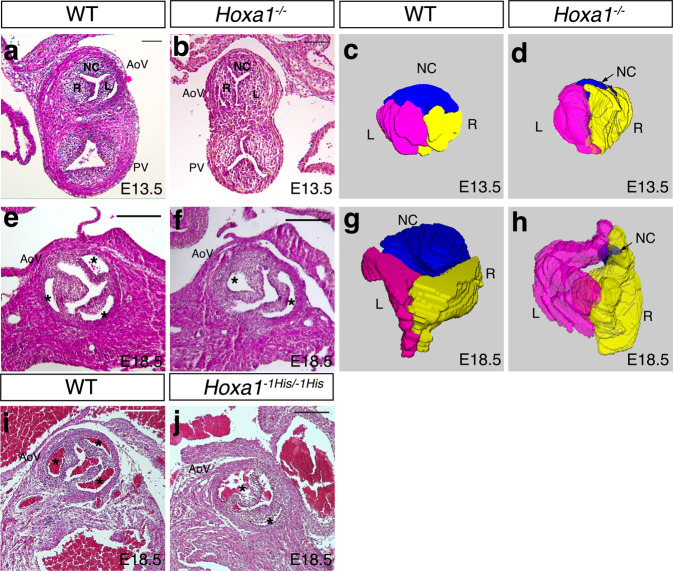
Fig. 4
a, b Hematoxylin and eosin (H&E) staining images showing representative transversal section of wild-type (WT) and Hoxa1−/− outflow tract at E13.5. Representative pictures from 3 independent hearts of each genotype. c, d Three-dimensional (3D) reconstructions of histological images at E13.5. A slight difference between WT (c) and Hoxa1−/− (d) aortic valve (AoV) structure is observed. Indeed, the non-coronary leaflet (NC) seems smaller in Hoxa1−/− when compared to wild-type embryos. e, f Cross-sectional H&E images through the aortic valve of WT (e) and Hoxa1−/− (f) embryos at E18.5. Representative pictures from 3 independent hearts of each genotype. Normal valve with three leaflets (asterisks) is observed in WT embryos (f), whereas bicuspid aortic valve is detected in the mutant (f). Asterisks indicate the aortic sinus. g, h 3D reconstruction of histological images at E18.5 showing three aortic valve leaflets in the WT embryo (g), whereas a persistent small non-coronary leaflet is seen in Hoxa1−/− embryos (h). i, j Cross-sectional H&E images through the aortic valve of WT (i) and Hoxa1−1His/−1His (j) embryos at E18.5. Representative pictures from 4 independent hearts of each genotype. Normal valve with three leaflets (asterisks) is observed in WT embryos (i), whereas bicuspid aortic valve is detected in the knock-in embryos (f). Left coronary (L; pink), right coronary (R; yellow), non-coronary (NC; blue) leaflets; PV, pulmonary valve. Scale bars: 100 μm (a, b); 200 μm (e, f, I, j).