
|
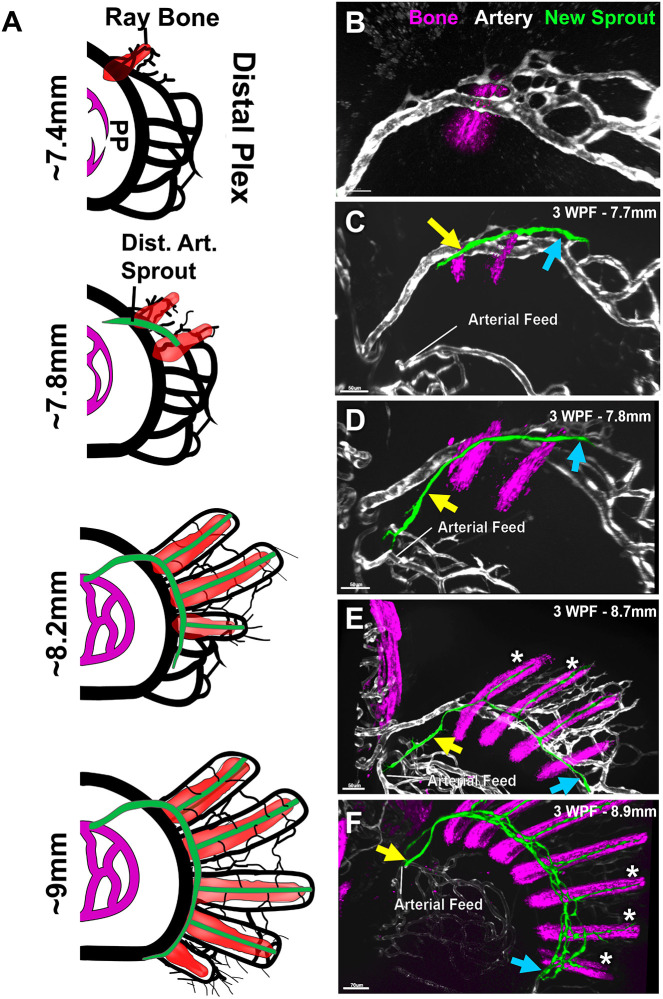
Fig. 6
The formation of the distal fin-ray arterial network begins as a sprout from the distal venous plexus. (A) Schematic of the relationship between the developing fin-ray bones (red) and developing fin-ray arterial network (green) that emerges from the pectoral fin distal vascular plexus (black) in ∼7.4-9.0 mm fish. (B-F) Confocal images of the pectoral fin distal vascular plexuses of separate 3-week-old Tg(Ola.Sp7:mCherryEco.NfsB)pd46, Tg(kdrl:egfp)s843 double-transgenic zebrafish of different sizes, with Tg(Ola.Sp7:mCherryEco.NfsB)pd46-positive condensing bone in magenta and Tg(kdrl:egfp)s843-positive vessels in gray, except for the emerging fin-ray arterial network which is false-colored in green. Larvae shown are 7.4 mm, (B), 7.7 mm (C), 7.8 mm (D), 8.7 mm (E) and 8.9 mm (F) in length. Yellow and blue arrows indicate the proximal/dorsal and distal/ventral ends, respectively, of the developing fin-ray arterial vascular network. White asterisks indicate developing fin-ray arteries. Images shown are representative of data collected from six separate larvae. Dist. Art., distal arterial sprout; Distal Plex, distal plexus; PP, proximal plexus; WPF, weeks post-fertilization. Scale bars: 50 μm (B-E); 70 μm (F).