
Fig. 9
- ID
- ZDB-IMAGE-230226-48
- Publication
- Vorontsova et al., 2022 - In vivo macromolecular crowding is differentially modulated by aquaporin 0 in zebrafish lens: Insights from a nanoenvironment sensor and spectral imaging
- All Figures
- Figures for Vorontsova et al., 2022
|
Fig. 9
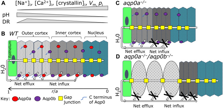
(A) Summary of physiological changes: intracellular Na+ ([Na+]i), Ca2+ ([Ca2+]i), crystallin concentrations ([crystallin]i), plasma membrane voltage potential (Vm), lens hydrostatic pressure (pi), and intracellular pH are shown as relative changes by gray bars, which occur from the outer cortex to the nucleus in a mature lens correlating to lens regions in (B). These changes determine its regional macromolecular crowding and, thus, DR. DR peaks in the outer cortex in mature lenses and decreases in the lens nucleus, indicating that the lowest macromolecular crowding and the highest water activity is in the outer lens cortex and lowest water activity is in the highly crowded lens nucleus. (B) Diagram of a mature equatorial lens fiber cell stack from epithelium (r/a = 1) to the center of the lens nucleus (r/a = 0) with direction of H20 flow shown on the basis of previously published work. (r is the distance from lens center and a is the lens radius). Aqp0a (red) and Aqp0b (purple) localize to both broad and narrow fiber cell membranes. Analysis of DR in aqp0a−/− (C) and aqp0a−/−/aqp0b−/− (D) lenses shows that both Aqp0a and Aqp0b facilitate osmotic water efflux in the outer cortex, while only Aqp0a facilitates water influx into fiber cells in the inner cortex (B). (C) Loss of Aqp0a results in a net loss of water influx, but Aqp0b is still able to facilitate efflux, resulting in net loss of water leading to increased macromolecular crowding and lower DR in the outer and inner cortex. (D) In double aqp0a−/−/aqp0b−/− mutants, both the H20 influx and efflux pathways are disrupted. This water loss results in a more crowded environment in the inner cortex with lower crowding in the swollen cells of the outer cortex.