
|
Figure 6
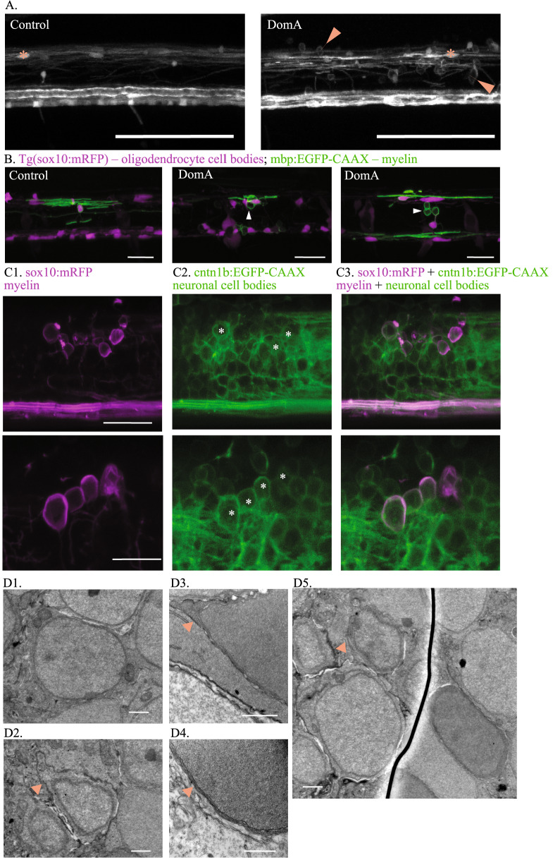
DomA exposure at 2 dpf leads to the formation of aberrant circular profiles that may be ectopically myelinated neuronal cell bodies. (A) Tg(mbp:EGFP) × Tg(mbp:EGFP-CAAX) double transgenic line labels both oligodendrocyte cell bodies and myelin sheaths. Asterisks mark oligodendrocyte cell bodies. Arrows label circular myelin membranes, which are commonly found in DomA-exposed larvae and rarely observed in controls. Scale bar = 100 μm. (B) Representative images of mosaically labeled oligodendrocytes in DomA-exposed (2 dpf) and control fish following 1–4 cell injections of the reporter construct, mbp:EGFP-CAAX into Tg(sox10:RFP) background, imaged at 4 dpf. Oligodendrocyte cell bodies are labeled in red. Arrows mark circular myelin membranes, which are outlined in green. These were the same fish used in Fig. 3, but with the red channel present to show the oligodendrocyte cell body. Scale bar = 25 μm. (C) Tg(sox10:mRFP) × Tg(cntn1b:EGFP-CAAX) double transgenic line imaged at 5 dpf. mRFP labels myelin sheaths and oligodendrocyte membranes. EGFP-CAAX labels the membrane of subpopulations of spinal cord neurons. Two examples of DomA-exposed double transgenic fish. Circular myelin membranes labeled in RFP (false colored magenta) (C1), neuronal cell bodies outlined in EGFP (C2). Images were then merged (C3). Asterisks mark neuronal cell bodies that are potentially associated with circular myelin membranes in the imaging plane. Top image scale bar = 25 μm., bottom image scale bar = 20 μm (D) Scanning electron micrograph of neuronal cell bodies in the spinal cords of Tg(mbp:EGFP-CAAX) fish. (D1) Example neuronal cell body in a control fish shows no evidence of myelin surrounding it. (D2–D5) Example neuronal cell bodies in DomA-exposed fish that may be wrapped by myelin. Arrows point to the putative myelin that surrounds the cell body. (n = 1 control, n = 1 DomA-exposed) Scale bar = 1 μm.