
|
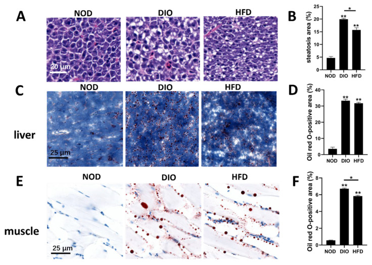
Figure 3
Characterization of hepatic steatosis and ectopic accumulation of lipid droplets in the liver and muscle. (A) Livers of zebrafish in NOD, DIO and HFD groups were analyzed with hematoxylin and eosin staining. (B) Quantitative analysis of the area of hepatic steatosis in zebrafish. Steatosis of three NOD, three HFD and three DIO across the liver were determined using the ImageJ software. Values are means ± SEM. **, p < 0.01. (C,E) Livers and muscles of zebrafish in NOD, DIO and HFD groups were analyzed with Oil Red O. (D,F) Quantitative analysis of the area of lipid droplets in liver and muscle. The areas of lipid droplets in the liver and muscle of three NOD, three HFD and three DIO were determined using the ImageJ software. Values are means ± SEM. *, p < 0.05. **, p < 0.01.