
|
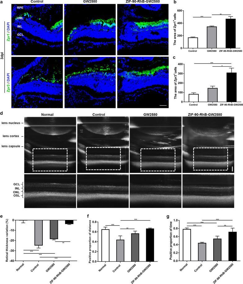
Fig. 7
The expression of photoreceptor markers, retinal structure, and optomotor response following treatment with ZIF-90-RhB-GW2580. a Zpr1 and Zpr3 immunofluorescence staining in sections taken from retinas in the control, GW2580 and ZIF-90-RhB-GW2580 groups at 3 days post-lesion (dpl). b, c Quantification of the fluorescence areas of b Zpr1- and c Zpr3-positive cells (ANOVA; *P < 0.05, **P < 0.01, ***P < 0.001). d Images of PS-OCT in retinas from the normal (unlesioned and uninjected), control, GW2580 and ZIF-90-RhB-GW2580 groups at 4 dpl. e Quantitative analysis of the retinal thickness variation (ANOVA; **P < 0.01, ***P < 0.001). f, g The statistical analyses of the f positive proportion of distance and g positive proportion of time at 4 dpl (ANOVA; **P < 0.01, ***P < 0.001). Scale bars in a: 20 μm; d: 100 μm. RPE, retinal pigment epithelium; ONL outer nuclear layer; INL inner nuclear layer; GCL ganglion cell layer; OSL outer segment layer