
|
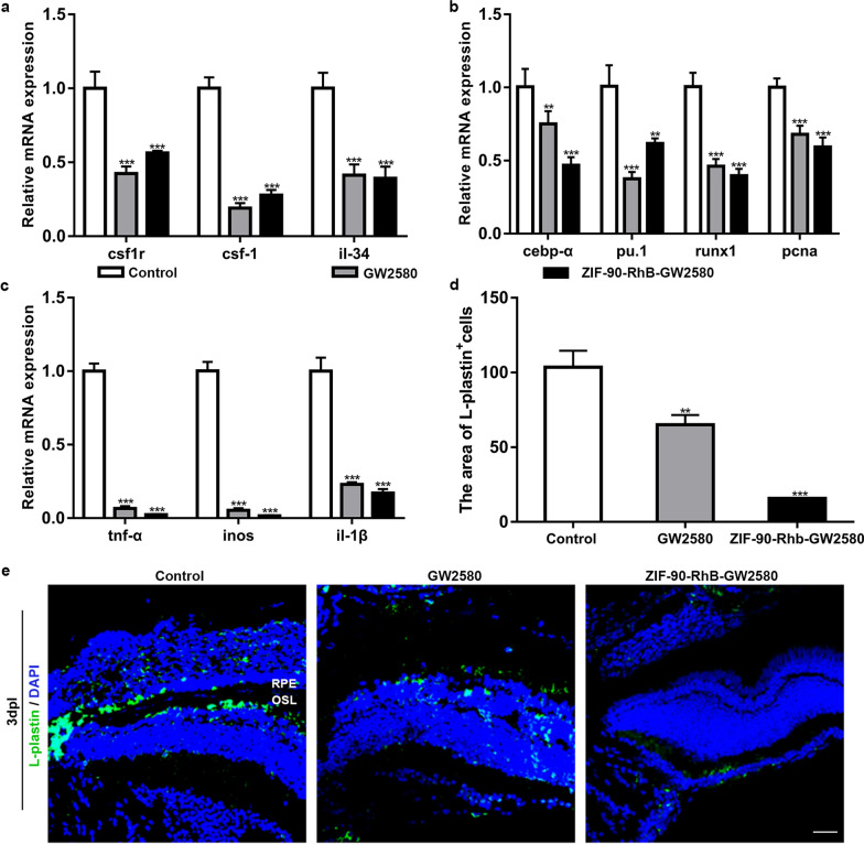
Fig. 6
The inhibited microglial activation and the expression of inflammatory factors following ZIF-90-RhB-GW2580 treatment. a qRT‒PCR analysis of the expression levels of csf1r, csf-1 and il-34 in the control, GW2580 and ZIF-90-RhB-GW2580 groups at 3 days post-lesion (dpl) (ANOVA; ***P < 0.001). b, c The relative expression levels of b microglial proliferation-related markers (cebp-α, pu.1, runx1 and pcna) and c proinflammatory factors (tnf-α, inos and il-1β) at 3 dpl (ANOVA; **P < 0.01, ***P < 0.001). d Quantification of the fluorescence area of L-plastin-positive cells in sections taken from retinas in three groups at 3 dpl (ANOVA; **P < 0.01, ***P < 0.001). e Immunofluorescence images of L-plastin at 3 dpl. Scale bar in e: 20 μm. RPE retinal pigment epithelium; OSL outer segment layer