
Image
|
Figure Caption
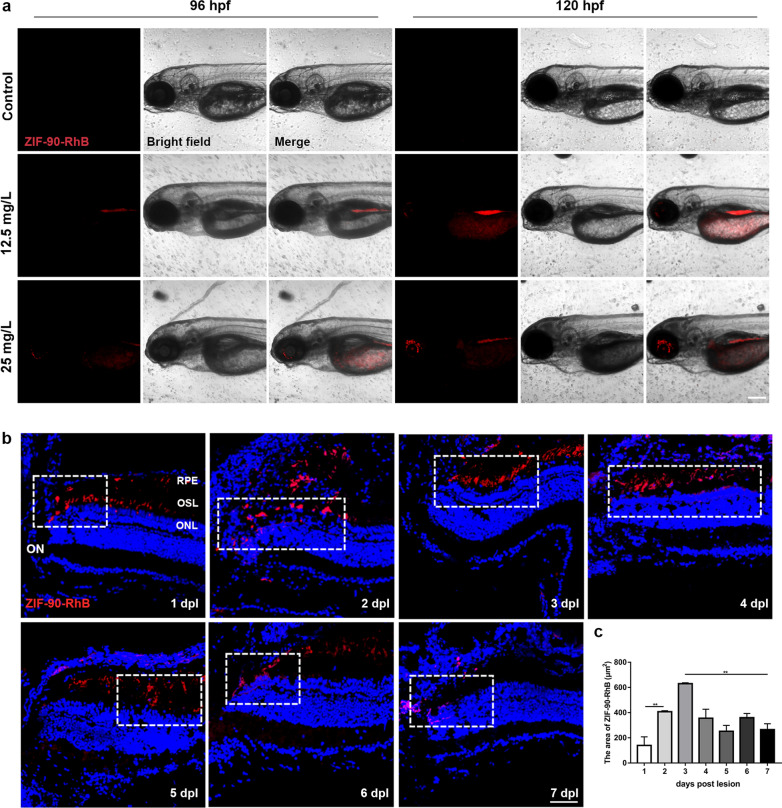
Fig. 4
The distribution of ZIF-90-RhB in larval zebrafish and light-lesioned retina. a Bright field and fluorescence images of larval zebrafish following ZIF-90-RhB exposure at 96 and 120 hpf. b Time-lapse localization of ZIF-90-RhB in sections taken from light-lesioned retinas from 1 to 7 days post-lesion (dpl). The lesion sites are indicated by the dotted rectangles. c Quantitative analysis of the fluorescence area (ANOVA; **P < 0.01). Scale bars in a: 500 μm; b: 20 μm. RPE retinal pigment epithelium; OSL outer segment layer; ONL outer nuclear layer; ON optic nerve
Acknowledgments
This image is the copyrighted work of the attributed author or publisher, and
ZFIN has permission only to display this image to its users.
Additional permissions should be obtained from the applicable author or publisher of the image.
Full text @ J Nanobiotechnology