
Image
|
Figure Caption
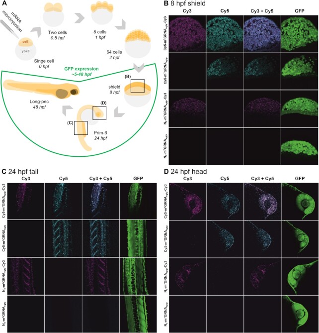
Fig. 6
Microinjection of zebrafish embryos with modified mRNA. (A) Schematic representation of the experimental set-up and injection of mRNA in the course of zebrafish development during the first 48 h post fertilization (hpf). (B–D) Confocal microscopy images of embryos injected with 300 pg of N3-m7GRNAegfp, N3-m7GRNAegfp-Cy3, Cy5-m7GRNAegfp or Cy5-m7GRNAegfp-Cy3 mRNA captured at 8 hpf (B) or 24 hpf in tail (C) or head (D) sections. Bright field and GFP fluorescence images of whole embryos are presented in Supplementary Figure S24.
Acknowledgments
This image is the copyrighted work of the attributed author or publisher, and
ZFIN has permission only to display this image to its users.
Additional permissions should be obtained from the applicable author or publisher of the image.
Full text @ Nucleic Acids Res.