
|
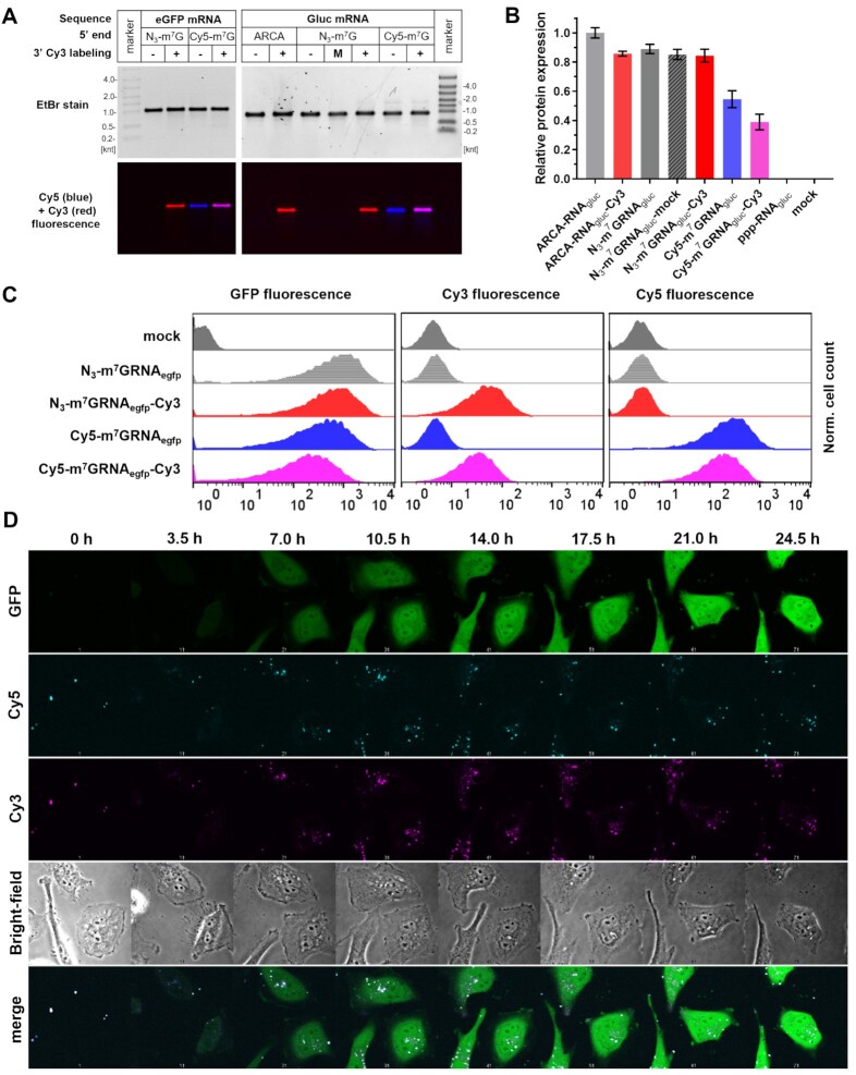
Fig. 5
Fluorescent mRNA transfection into HeLa cells. (A) Electrophoretic resolution of HPLC-purified mRNAs encoding eGFP (N3-m7GRNAegfp, N3-m7GRNAegfp-Cy3, Cy5-m7GRNAegfp and Cy5-m7GRNAegfp-Cy3) or Gaussia luciferase (ARCA-RNAgluc, ARCA-RNAgluc-Cy3, N3-m7GRNAgluc, N3-m7GRNAgluc-mock, N3-m7GRNAgluc-Cy3, Cy5-m7GRNAgluc and Cy5-m7GRNAgluc-Cy3) used for HeLa transfections. (B) Relative expression of Gaussia luciferase after mRNA transfection as a function of the presence of 3′ Cy3 modification and 5′ cap structure. (C) Flow cytometry readouts after transfection of fluorescent mRNA encoding eGFP. (D) Time-lapse microscopy images of HeLa cells transfected with Cy5-m7GRNAegfp-Cy3 mRNA. Time scale set for 0 h at the beginning of recording of the images (1 h after transfection start).