
|
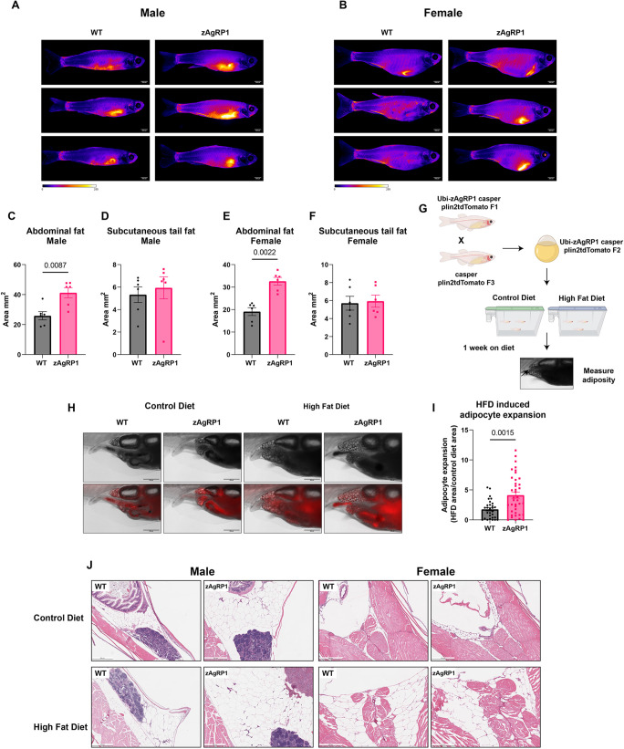
Fig. 2. Agrp overexpression increases visceral adiposity and susceptibility to high-fat diet (HFD). (A,B) Representative images of male (A) and female (B) BODIPY-stained fish. (C-F) Quantification of visceral abdominal (C,E) and subcutaneous (D,F) fat depots from BODIPY-stained male (C,D) and female (E,F) fish. n=6 fish per condition over three biological replicates. (G) Schematic of Plin2tdTomato HFD experiment. 21 dpf zAgRP1 or wild-type Plin2tdTomato fish were put on either a control diet or HFD for 1 week, and visceral adiposity was measured. Created with Biorender.com. (H) Representative images of Plin2tdTomato fish. (I) Quantification of adipocyte expansion. HFD-induced adipocyte expansion was calculated by taking the ratio of the area of adipocyte tissue on the control diet versus HFD for each genotype. n≥30 fish per genotype across three biological replicates. (J) Histology of cross-sections of male and female adult quad zAgRP1 fish or wild-type controls on an HFD or control diet for 3 months. Fish were fixed in PFA, sectioned and stained with H&E. Two fish per condition were sent for sectioning. Mann–Whitney test. Scale bars: 2.5 mm (A,B), 500 μm (H,J).