
Image
|
Figure Caption
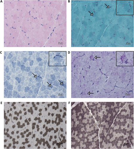
Fig. 2
Muscle biopsy finding based on histological examination: (A) Hematoxylin and eosin (H&E) stain: musculature seen transversely and obliquely, preserved fascicle structure; perimysial connective tissue proliferation, no fat cell proliferation; no lymphohistiocytic cell infiltrates; muscle fibers polygonal, physiological fiber size variability, no fiber fragmentation or nuclear clumps; predominantly peripheral nuclei (centrally located nuclei <3%). No cell necrosis or basophil regenerating fibers; no ragged red fibers; no intracellular glycogen or lipid proliferation. (B) Modified Gomori stain shows occasionally the enrichment of mitochondria subsarcolemmally (white arrows). (C) Nicotinamide adenine dinucleotide hydrogen (NADH) stain shows increased subsarcolemmal reactivity in a minority of fibers (white arrows). (D) Periodic acid–Schiff (PAS) stain shows increased sarcoplasmic build-up on a minority of diseased muscle fibers (white arrows). (E, F) Adenosine triphosphatase (ATPase) stain (at pH 4.3 and 9.4) shows fiber-type disproportion (increased abundance of type 2 fibers). Insets show areas of interest in greater detail.
Acknowledgments
This image is the copyrighted work of the attributed author or publisher, and
ZFIN has permission only to display this image to its users.
Additional permissions should be obtained from the applicable author or publisher of the image.
Full text @ J. Pathol.