
|
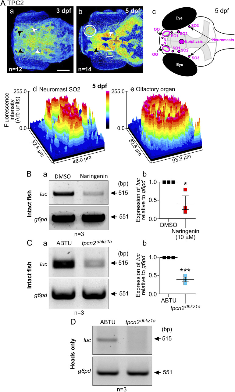
FIG 6 Effect of pharmacologically mediated inhibition of TPC2 activity or CRISPR/Cas9-mediated tpcn2 knockout on PEG-pseudovirus entry in zebrafish larvae. (A) (a and b) Immunolabeling analysis of TPC2 localization in the head at 3 dpf and 5 dpf (viewed from a dorsal orientation) showing distinct expression in the olfactory organ (black arrowheads), neuromasts (white arrowheads), and epiphysis (brown arrowheads). (c) Schematic showing a dorsal view of the head of a zebrafish larva at 5 dpf indicating the position of the olfactory organs (OO), nasal (N) and supraorbital (SO1 to SO3) neuromasts, and epiphysis. (d and e) Surface plots showing the fluorescence intensity in neuromast SO2 and the olfactory organ, respectively. Bar, 100 μm. (B) RT-PCR analysis of cDNA obtained at 2 dpf from intact wild-type embryos that were pretreated with naringenin or DMSO (control) prior to exposure to live PEG-pseudovirus for 48 h. (a) Representative electrophoresis gel showing luc expression and (b) quantification of the relative expression of luc in the naringenin group compared to the DMSO group. (C) RT-PCR analysis of cDNA obtained from intact wild-type and tpcn2dhkz1a embryos, which were exposed to live PEG-pseudovirus at 2 dpf for 48 h. (a) Representative electrophoresis gel showing luc expression and (b) quantification of the relative expression of luc in the tpcn2 mutant compared to the wild-type control. (D) Representative electrophoresis gel of luc expression from RT-PCR analysis of cDNA extracted from the excised heads alone of wild-type and tpcn2dhkz1a embryos that had been exposed to live PEG-pseudovirus at 2 dpf for 48 h.