
Image
|
Figure Caption
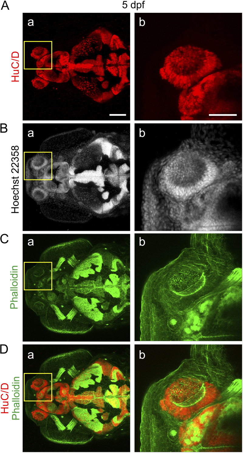
FIG 4 Localization of neurons in the olfactory organ of larvae at 5 dpf. Representative (n = 7) dorsal views of a larva (A) immunolabeled with anti-HuC/D, to label neuronal nuclei, and then stained with (B) Hoechst 33258 and (C) fluorescent phalloidin, to label the nuclei and F-actin, respectively. (D) Merged HuC/D and phalloidin images. The regions bounded by the yellow squares in panels a are shown at higher magnification in panels b. Bars, 100 μm (panels a) and 50 μm (panels b).
Acknowledgments
This image is the copyrighted work of the attributed author or publisher, and
ZFIN has permission only to display this image to its users.
Additional permissions should be obtained from the applicable author or publisher of the image.
Full text @ J. Virol.