
|
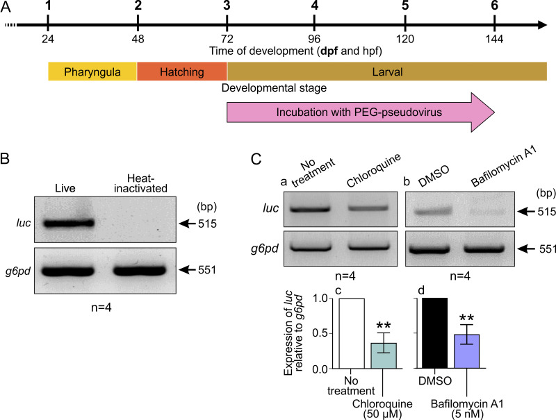
FIG 2 Effect of incubating ABTU wild-type embryos with PEG-pseudovirus at ~3 dpf for 72 h. (A) Schematic to show when embryos were incubated with PEG-pseudovirus. (B) Representative (n = 4) gel electrophoresis image showing RT-PCR results from cDNA extracted from whole ABTU larvae exposed to live or heat-inactivated PEG-pseudovirus. (C) (a and b) Representative gel electrophoresis images and (c and d) bar charts of the RT-PCR results from cDNA extracted from whole ABTU larvae exposed to live PEG-pseudovirus and treated with (a and c) 50 μM chloroquine or (b and d) 50 nM bafilomycin A1 (n = 4 for each). The level of expression of luciferase (luc) mRNA was determined against that of glucose-6-phosphate dehydrogenase (g6pd) mRNA, and the gene expression level following treatment with chloroquine or bafilomycin A1 was measured relative to the respective untreated or DMSO-treated controls. Values are means and standard errors of the means (SEM) from 4 experiments. A 2-sample t test was used to calculate statistical significance. **, P < 0.01.