
|
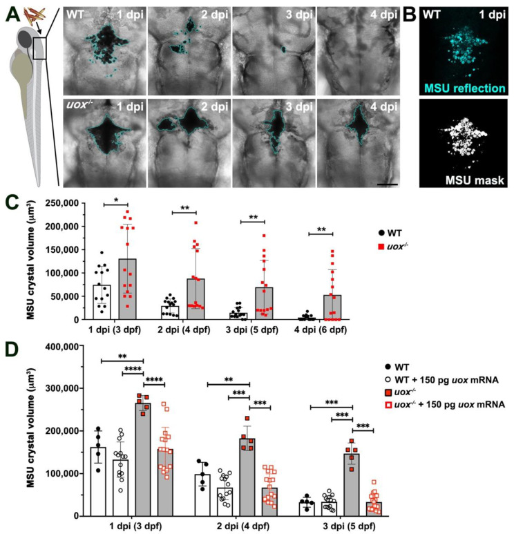
Figure 4
Microinjected MSU crystals persist in uox−/− larvae. (A) Representative confocal imaging (transmitted light views, Z-projections) of WT and uox−/− larvae at 1, 2, 3 and 4 days post-injection (dpi) with MSU crystals into the hindbrain ventricle (injected at 2 days post fertilization (dpf)). A cyan border outlines MSU crystals. (B) A representative reflection image of MSU crystals in a WT larva at 1 dpi (same larva as in (A)), pseudo-coloured in cyan. The lower panel shows a standard deviation Z–projection of the binary mask in black and white. (C) Quantification of MSU crystals (μm3), as detected in (B), within individual WT (white bars) and uox−/− (grey bars) larvae at 1, 2, 3 and 4 dpi (3, 4, 5 and 6 dpf, n = 15 larvae per group). Statistical significance determined using multiple unpaired Student’s t-tests with Holm–Sidak correction. (D) Quantification of MSU crystals (μm3) within individual WT (white bars) and uox−/− (grey bars) larvae at 1, 2 and 3 dpi (3, 4 and 5 dpf) and within WT (white bars) and uox−/− (grey bars) larvae injected with 150 pg uox mRNA (n= 5 and 15 larvae per group for the non-mRNA-injected and mRNA-injected groups, respectively). Statistical significance determined using a 2-way ANOVA and Tukey’s multiple comparisons test. Error bars in (C,D) represent means ± SDs. * p < 0.05, ** p < 0.01, *** p < 0.001 and **** p < 0.0001. Scale bar 100 µm.