
|
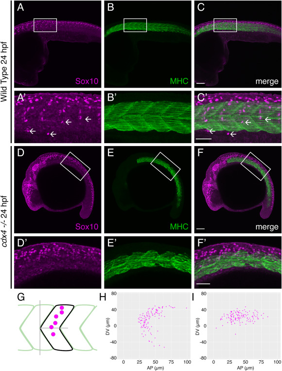
Fig. 4 cdx4 mutants show impaired segmental migration of trunk NC cells. A-C. Lateral view of 24 hpf WT embryo labeled with anti-Sox10 (A, NC cells, magenta) and anti-Myosin Heavy Chain (B, somites, green) antibodies reveal segmental migration pathways of trunk NC cells, scale bar = 100 μm. A’-C’. High magnification images of boxed area from A-C (arrows = individual segmental chains), scale bar = 50 μm. D-F. Lateral view of 24 hpf cdx4ch107−/- embryo labeled with anti-Sox10 (D, NC cells, magenta) and anti-Myosin Heavy Chain (E, somites, green) antibodies reveal defects in trunk NC cell migration. Scale bar = 100 μm. D’-F’. High magnification images of boxed area from D-F, scale bar = 50 μm. G. Schematic representing approach for mapping the position of trunk NC cells relative to the adjacent somite; analysis was performed at the level of somites 5–10. H, I. Plots of migrating trunk NC cells relative to the adjacent somite in WT embryos (H) and cdx4ch107−/- embryos (I). Data from 18 total segments were superimposed on each plot.
Reprinted from Developmental Biology, 480, Rocha, M., Kushkowski, E., Schnirman, R., Booth, C., Singh, N., Beadell, A., Prince, V.E., Zebrafish Cdx4 regulates neural crest cell specification and migratory behaviors in the posterior body, 25-38, Copyright (2021) with permission from Elsevier. Full text @ Dev. Biol.