
Fig. 6
- ID
- ZDB-IMAGE-221119-32
- Antibodies
- Publication
- Coffey et al., 2021 - Lysosomal Function Impacts the Skeletal Muscle Extracellular Matrix
- All Figures
- Figures for Coffey et al., 2021
|
Fig. 6
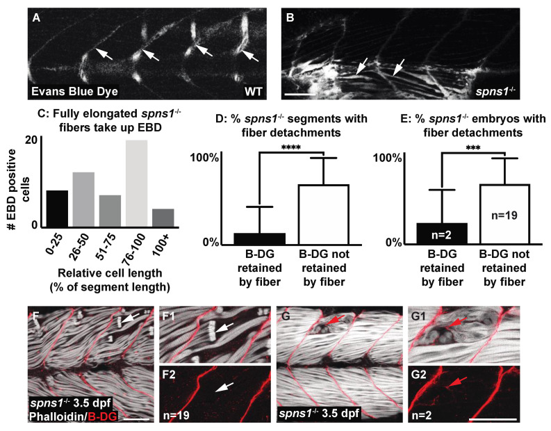
Muscle fiber failure is due to sarcolemmal integrity failure in spns1−/− 3.5 dpf larvae. (A,B,F,G) Anterior left, dorsal top, side-mounted 3.5 dpf larvae. (A,B) WT and spns1−/− 3.5 dpf larvae injected with 1% Evans Blue Dye (EBD). (A,B) White arrows indicate the presence of EBD. (A) In WT larvae EBD remains in the bloodstream but in (B) spns1−/− larvae EBD penetrates full length muscle fibers, indicating membrane damage. (C) Quantification of relative cell length of EBD-penetrated myofibers in spns1−/− larvae shows that fully elongated fibers in spns1−/− larva take up EBD. (D) Percentage of segments with beta-dystroglycan containing fibers and beta-dystroglycan negative fibers in spns1−/− larvae. Note that beta-dystroglycan remains at the MTJ the majority of the time. (E) Percentage of spns1−/− larvae with beta-dystroglycan containing fibers and beta-dystroglycan negative fibers. *** p < 0.001, **** p < 0.0001. (F–G2) Phalloidin (white) and beta-dystroglycan staining (red) of 3.5 dpf spns1−/− larvae. (F,F1,G,G1) Merged phalloidin and beta-dystroglycan channels and (F2,G2) beta-dystroglycan single channel. (F–F2) White arrows point to retracted fibers that do not contain beta-dystroglycan at their detached ends. (G–G2) Red arrows point to retracted fibers that contain beta-dystroglycan at their detached ends. Scale bars 50 µm.